Researchers from Tohuku University in Japan have developed a method of using x-ray elastography to produce detailed images of soft tissues with much greater resolution than ultrasound or MRI.
This greater resolution for the field of elastography—a non-invasive method of medical imaging that investigates the stiffness and elasticity of soft tissue—should allow healthcare professionals to identify much smaller and deeper tissue problems, such as lesions, than they can with ultrasound or MRI, the two main types of elastography used currently.
Although previous studies have suggested such x-ray elastography is possible in principle, this is the first time that any real-world visualization of stiffness using the concept has been demonstrated.
“This greater precision doesn’t just mean identification of much smaller or deeper lesions,” said lead researcher Wataru Yashiro, an associate professor from the Institute of Multidisciplinary Research for Advanced Materials (IMRAM) of Tohoku University, “but, importantly for patients, because smaller lesions can be newer ones, potentially also much earlier on in a disease or condition.”
The next step is to further develop the technique to produce 3D visualizations, and ultimately the researchers want to manufacture x-ray elastography medical diagnostic equipment.
Read more from Tohuku University and find the paper at Applied Physics Express.
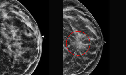
Featured image: Maps of stiffness (storage modulus) in uniform-concentration sample (left) and sample with harder inclusion (right) (sample: polyacrylamide gel). It can be seen that harder inclusion is clearly visible in spite that its concentration is only slightly different from the surrounding matrix. Note that such a slight difference cannot be discerned by typical X-ray radiography for medical diagnostics. Credit: Wataru Yashiro, the Institute of Multidisciplinary Research for Advanced Materials (IMRAM), Tohoku University.