A new approach to functional bone imaging has established that bone metabolism is abnormally elevated in patients with knee osteoarthritis. This physiological information provides a new functional measure to help assess degeneration of the knee joint. The research was presented at the Society of Nuclear Medicine and Molecular Imaging 2020 Virtual Annual Meeting, July 11-14.
Osteoarthritis, the most common joint disorder in the United States, affects more than 32.5 million adults. It is marked by degradation and loss of soft tissues, such as cartilage, and development of bone marrow lesions and osteophytes. Osteoarthritis occurs most frequently in the hands, hips and knees and results in reduced productivity and quality of life.
“Osteoarthritis is not well understood, in part because we lack the tools to objectively evaluate early and reversible changes in key tissues,” notes Lauren Watkins, MS, a researcher at the Stanford University Imaging of Musculoskeletal Function Group in Stanford, California. “While many MRI methods have been developed for assessment of early degenerative changes in cartilage, functional imaging of bone in the joint remains a major challenge.”
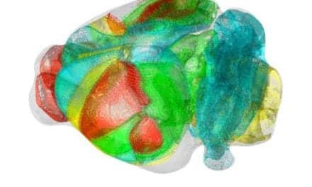
To evaluate the relationship between structural and physiological changes in knee osteoarthritis, researchers utilized PET/MRI with 18F NaF to image both knees of 12 subjects. Five subjects were scanned twice, with at least five days between visits, to assess repeatability of the technique. MRI Osteoarthritis Knee Score (MOAKS) assessment of each knee was performed by a trained musculoskeletal radiologist, and dynamic PET data were used to calculate the rates of bone perfusion, tissue clearance and mineralization, as well as tracer extraction fraction and total bone uptake rate. Kinetic modeling was performed for regions of interest representing the subchondral bone of the patella, medial and lateral tibia, and anterior, central, and posterior regions of the medial and lateral femur.
The knees with MOAKS findings were divided into regions of cartilage loss, bone marrow lesions and osteophytes and were analyzed along with the kinetic parameters derived from PET data. Abnormal bone metabolism in regions with bone marrow lesions, osteophytes, and adjacent cartilage lesions was found to be strongly associated with greater bone perfusion rates as compared to bone that appeared normal on MRI. Additionally, strong spatial relationships between bone metabolic abnormalities and changes in overlying cartilage were noted.
“These findings show the utility and potential of PET imaging to study the role of bone physiology in degenerative joint disease,” says Watkins. “This knowledge may help us understand the order of events leading to structural and functional degeneration of the knee. Further, this will help us to develop and quickly evaluate new interventions that target specific metabolic pathways to give us the best chance to slow or arrest the onset and progression of osteoarthritis.”