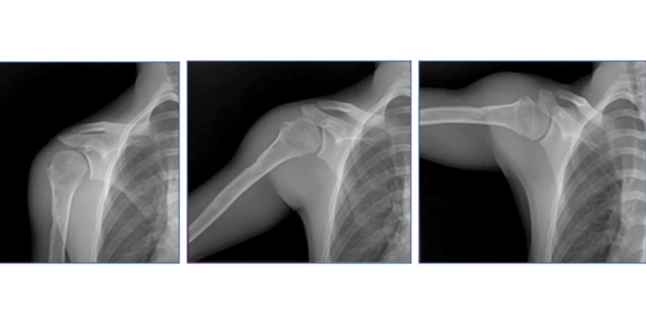
Researchers used low-dose DDR cineloops to measure scapulohumeral rhythm following anatomic and reverse total shoulder arthroplasty, highlighting the modality’s potential role in functional imaging and post-operative assessment.
Researchers at Emory Healthcare have demonstrated how dynamic digital radiography (DDR) can be used to measure functional outcomes following total shoulder arthroplasty, providing new insights into how different surgical techniques restore shoulder biomechanics.
The study, published in the Journal of Shoulder and Elbow Surgery and led by Eric R Wagner, MD, compared two established treatment options for cuff-intact glenohumeral osteoarthritis: anatomic total shoulder arthroplasty (aTSA) and reverse total shoulder arthroplasty (rTSA).
Using DDR to image shoulders while in motion, the research team evaluated scapular contributions to shoulder elevation in patients after both surgical procedures. The technology captures a series of static images in quick succession to create detailed visualizations of joint movement over time.
Similar Results Between Surgical Techniques
In a retrospective analysis of 71 shoulders treated with either arthroplasty technique compared to 32 normal controls, researchers found similar scapulohumeral rhythm (SHR) for both aTSA and rTSA procedures, though neither achieved normal biomechanics.
The study revealed that while the rTSA group had fairly constant SHR throughout shoulder elevation, aTSAs showed higher SHR in the second half of shoulder motion compared to the first half, suggesting greater glenohumeral involvement as abduction progresses.
“While our study reports a significant improvement in scapular motion post-arthroplasty using either aTSA or rTSA, native scapulohumeral biomechanics is not restored by either surgical technique,” says Sameer R. Khawaja, MD, orthopaedic surgery resident at Baylor College of Medicine, in a release.
Clinical Applications for Pre-Surgical Planning
The findings may help inform pre-surgical planning regarding technique selection and implant positioning, particularly for younger active patients where restoring shoulder function and native kinematics may be more important.
“Our goal is to help inform pre-surgical planning regarding selection of technique and implant positioning, particularly for younger active patients where restoring shoulder function and native kinematics may be more desired,” says Khawaja in a release. “We are currently finalizing a study examining the correlation between scapular motion and patient outcomes using DDR.”
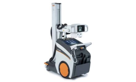
DDR is a low-dose X-ray imaging technique that generates cineloops enabling clinicians to visualize anatomical motion over time. The technology provides an alternative method for measuring joint function compared to traditional static imaging approaches.
“By using DDR to measure SHR and quantify the glenohumeral and scapulothoracic contributions to humerothoracic abduction, the Emory team has demonstrated a method to quantify functional outcome post-arthroplasty and laid the foundation for enhanced range of motion in future shoulder arthroplasties,” says John Sabol, PhD, clinical research manager at Konica Minolta Healthcare, which manufactures the DDR system used in the study, in a release.
The research addresses a gap in understanding how alterations in scapular motion affect clinical outcomes in patients with glenohumeral osteoarthritis following total shoulder arthroplasty. While it is known that patients have reduced scapulohumeral rhythm compared to healthy subjects after shoulder arthroplasty, it remained unclear whether the lower SHR relates to preoperative pathology or surgical intervention.
Photo caption: Dynamic digital radiography image
Photo credit: Konica Minolta Healthcare