A radiologist discusses the evolution from traditional CAD to modern AI, its impact on burnout, and the future of personalized risk assessment.
By Alyx Arnett
When breast radiologist Svati Singla Long, MD, began her career nearly 15 years ago, computer-aided detection (CAD) was the main tool available to help radiologists spot potential cancers on mammograms. Today, she says, artificial intelligence (AI) represents the next stage of that evolution—one that’s already changing how clinicians read images, manage workloads, and approach patient risk.
“Breast AI is best utilized in a collaborative way: using the technology as part of the radiologists’ toolset,” Long says. “The radiologists are still the ultimate expert responsible for the decision-making, but the AI has shown great promise in making us better at what we do.”
As the part-time medical director of the US division of Therapixel, a French software company specializing in AI applied to medical imaging, Long has had a front-row seat to AI’s growing role in breast imaging. She offers a look inside the technology’s impact today and its potential for the future.
From Rule-Based CAD to Learning Algorithms
The journey of computer assistance in mammography began long before the current AI boom. The first US Food and Drug Administration (FDA)-approved CAD system arrived in 1998, becoming widely adopted by the early 2000s. According to Long, this traditional CAD relied on predefined rules to flag patterns like shapes and densities for radiologist review.
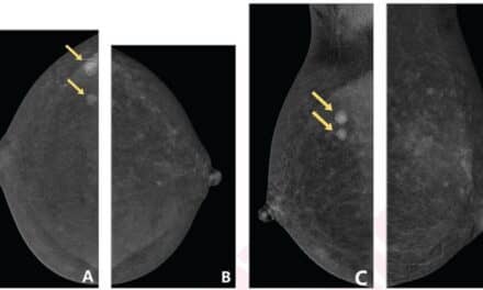
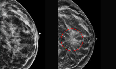
In contrast, the AI platforms that entered the market in the 2020s operate on a different principle. “AI cancer detection entered the arena with an adaptive algorithm that learns directly from thousands or millions of mammograms to help the radiologist identify possible cancer,” Long says.
These advanced systems can integrate 2D and 3D images along with prior studies to provide a more comprehensive analysis, aiming to enhance cancer detection and reduce human error.
A Tool to Enhance Confidence and Reduce Burnout
Beyond improving detection rates, Long says one of AI’s biggest benefits is its ability to ease the daily pressure on radiologists. The high volume and repetitive nature of screening mammography can lead to fatigue, but AI offers a valuable “second look” that can boost confidence and reduce stress.
“I can tell you that when I utilize AI to read, I do feel less burnt out,” Long says. “Having the AI simultaneously ‘read’ the mammogram with me provides an extra layer of confidence as I go through my list.”
That experience echoes what Therapixel found when conducting a reader study for FDA clearance of its AI tool, MammoScreen—83% of participating radiologists said they felt less burnout when using the system compared with reading without it.
Integrating AI into the Clinical Workflow
For any new technology to succeed in radiology, it has to fit naturally into the way clinicians already work, says Long. In breast imaging, that means providing real value without slowing down the reading process.
“You want a seamless integration that provides benefits without increasing the reading time,” Long says.
She adds that building trust in AI is just as important—and that comes through rigorous testing and clinical validation before the technology is introduced into practice. Once radiologists see the results for themselves, adoption tends to happen quickly.
“I’ve heard of some radiologists refusing to read without [AI] even after just a few days of using it in their clinic because they find it so helpful,” she says.
The Future: Personalized Risk Assessment
Looking ahead, Long is most excited about AI tools that can estimate a woman’s breast cancer risk directly from her mammogram images.
Traditional risk models depend largely on patient-reported details such as family history and reproductive factors. AI, she says, is starting to offer a more objective, personalized approach.
“Now with AI, we are seeing the technology calculate a risk based on each woman’s actual mammographic images,” Long says. This, she adds, is “paving the next step for true personalization of cancer risk, which will ultimately lead to early detection.”
By moving beyond self-reported data and focusing on tissue patterns visible on the mammogram, Long believes AI can help radiologists tailor screening recommendations and catch cancers earlier.
ID 42476235 | Breast Imaging © Zlikovec | Dreamstime.com