By Karen Appold
The Hospital of Central Connecticut (HOCC), New Britain, Conn, was the first medical facility in Connecticut to adopt VolparaDensity breast imaging software, designed to help overcome the limitations of mammography to detect cancer in women with dense breasts.
Research shows that mammography in women with dense breast tissue may be less than 40% to 70% accurate.1 For women with extremely dense breasts, there may be an even greater chance that cancer is being masked. Furthermore, these women’s risk of developing breast cancer may increase by four to six times over women without dense breasts.
Studies have shown that about 40% of women have breast tissue that is greater than 50% dense.2 This is influenced by genetics and being a younger age (because these women have more hormones). But some postmenopausal women still have greater than 50% breast tissue density.
Connecticut was the first state to adopt legislation that requires radiologists to notify patients if they see dense tissue on a mammogram and stress the importance of talking with their doctor about having additional imaging studies—such as a bilateral breast ultrasound.

Jean Weigert, MD, FACR, Hospital of Central Connecticut
Research in the early 2000s showed that with an additional bilateral breast ultrasound, an additional four cancers per 1,000 were discovered that weren’t seen on a mammogram.3 “For the most part, these were small, node negative cancers that most likely were easier to treat,” said Jean Weigert, MD, FACR, director of breast imaging, HOCC. Although studies have yet to confirm this, “one would think that finding cancer at an earlier stage would improve mortality.”
Assigning breast density can be very subjective, Weigert points out. “One radiologist may interpret a mammogram as being over 50% dense while another one may say it’s less than 50%.” Variables such as the technologist who performs the mammography? and the type of unit it is performed on play a role.
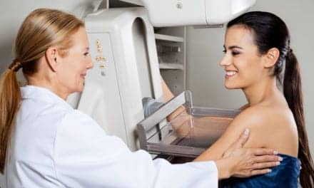
Just before the New Year, HOCC took action to take the guesswork out of determining breast density by adopting Wellington, New Zealand-based Matakina International’s VolparaDensity software. VolparaDensity calculates volumetrically the density of breast tissue on full field digital mammography. “The program analyzes the raw data from the digital images to calculate the volumetric density,” Weigert explained.
Weigert says HOCC adopted the software because “We don’t want to give ultrasounds to patients who don’t need them, but we do want to use it on those who do.” There is no additional charge to the patient to use the software.
While considering the software, Weigert thought it would improve HOCC’s ability to be consistent and accurate in determining breast tissue density. “I think referring doctors want to know that the right studies are being done for their patients and that they have a scientific basis for it,” she added.
VolparaDensity meets the requirements for designation of breast density according to the American College of Radiology’s Breast Imaging Reporting and Data System (BI-RADS) categories 1-4 by assigning a computer-derived value. “This value is objective and accurate, having been tested and cross calibrated from mammogram machine to machine as well as MRI-derived densities,” said Weigert.
Despite these advances, conducting bilateral breast ultrasound remains controversial. “It is not completely accepted in the imaging community,” Weigert said. More doctors need to be better educated.
According to data Weigert collected, the first year that ultrasounds were recommended, 8,000 women had them. In the second year, 10,000 women had them. “We found more than 3.2 additional cancers per 1,000 patients screened with bilateral breast ultrasound,” Weigert reported. “That is almost double the number of cancers you find with mammography. For the diagnosed women, it has been quite compelling.”4
Weigert says the timing in adopting VolparaDensity software revolved around having it approved by the Food and Drug Administration and having other imaging facilities use it first.
Weigert is optimistic that VolparaDensity will improve outcomes. “If a patient is on the border of having dense breast tissue–say between 45% and 55%—and the software determines the patient to be more than 50% dense, I will agree with that number.”
In addition, VolparaDensity can examine the raw data–such as how much the breast is compressed and the radiation dose. “These and other parameters may make us better mammographers,” Weigert said.
Like any new technology, there is a learning curve. “When we’ve pointed out a few problems, the company has been extremely aggressive with making sure they fix them right away,” she said.
###
REFERENCES
- Gottlieb S. Ultrasound plus mammography may detect more early cancers. BMJ. 2002;325(7366):678.
- Stomper PC, D’Souza DJ, DiNitto PA, Arredondo MA. Analysis of parenchymal density on mammograms in 1353 women 25-79 years old. AJR Am J Roentgenol. 1996;167:1261-5.
- Kolb TM, Lichy J, Newhouse JH. Comparison of the performance of screening mammography, physical examination, and breast US and evaluation of factors that influence them: an analysis of 27,825 patient evaluations. Radiology. 2002;225:165-175
- Weigert J, Steenbergen S. The Connecticut experiment: the role of ultrasound in the screening of women with dense breasts. Breast J. 2012;18(6):517-522.