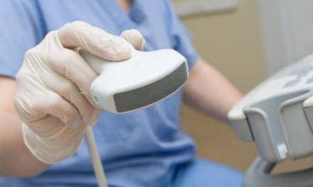
 When it comes to managing ultrasound images, there are two paths to follow — single-modality PACS and multimodality PACS.
Many radiology departments have adopted strategic goals to develop a filmless environment where providers accomplish diagnostic and reporting functions for all medical imaging procedures. Ultrasound images either can be managed using a modality-specific system or within a multimodality PACS (picture archiving and communications system), and the possible additional integration into HIS/RIS systems.
Healthcare systems are faced with decisions about which path to take. A niche single-modality PACS addresses the explicit requirements for dynamic ultrasound studies or the integration of those images into an existing enterprisewide multimodality PACS. Ultrasound equipment vendors have grappled with these issues for as long as they have networked equipment. Enterprisewide PACS vendors have begun to develop solutions to the unique image management problems presented by integrating ultrasound into their systems.
Critical issues
Many ultrasound studies include color segments to differentiate various anatomical structures and the use of cine or dynamic image clips to incorporate motion into the diagnostic features of a study.
Dean Terry, general manager of Acuson Corp.’s KinetDx division (Mountain View, California), uses a freeway metaphor to explain the importance of motion in an imaging study. A driver on an interstate sees the cars in front or in back of his car and is aware of cars on the on-ramp, off-ramp, overall traffic patterns and so on. A dynamic cine-clip in an ultrasound study offers data not possible in a still-frame picture.
“The feedback we get is that adding clips into a protocol for a general abdominal exam gives a higher level of confidence in diagnosis,” Terry explains.
Acuson’s KinetDx PACS is designed for cardiology, radiology, vascular labs and obstetrics, and designed and built from the ground up to perform in an echocardiology lab. Ultrasound exams offer 3D image information that is not available with all imaging modalities.
The DICOM standard for image compression, JPEG multiframe objects, provides necessary support for electronic storage and retrieval functions for ultrasound cine-clips on a multimodality PACS.
“Most enterprisewide PACS vendors don’t support DICOM, JPEG multiframe objects,” says Terry, “but they’re all committed to supporting that in the next release of their software. They’re working with us and we’re proactively working with them to test and validate and assure that their implementation performs reliably with a high degree of performance.”
Compression is required, because ultrasound image files are huge and require a long period of time to transmit unless the image is compressed.
“One cine-clip that is 4 to 5 seconds long could be as much as 100 megabytes of information,” adds Dana Driver, director of marketing and ultrasound at A.L.I. Technologies Inc. (Vancouver, British Columbia, Canada). “If we compress that information, not only is there better speed of transmission, but it also compresses to replay it in less time.”
The JPEG Lossy compression by ALI offers a 30:1 compression that is FDA approved. In a lossy compression, some image data is removed from the exam, but there is “no significant loss from the diagnostic information,” says Driver. “It provides an excellent ability to archive by a factor of five times as much information as if you were archiving at a 2:1 ratio.”
One of the challenges facing multimodality PACS vendors that need to integrate ultrasound is how those images are stored for future retrieval.
Bruce R. Parker, M.D., chairman of the department of diagnostic imaging at Texas Children’s Hospital and professor of radiology and pediatrics at Baylor College of Medicine (Houston), is using an Acuson ultrasound system, which is integrated into an Agfa Impax system. He explains that their cine-clips are usually about six seconds long. When they transfer a clip to their multimodality PACS it makes it available throughout the enterprise. An initial problem in transferring images to AGFA’s Unix-based release 3.5 has been resolved by a workaround, but smooth integration with Agfa’s NT-based release 4.0 is expected.
Stand-alone ultrasound systems
Stand-alone, modality-specific ultrasound systems offer features for image capture, storage, retrieval and distribution across computer networks. There are advantages and disadvantages to adopting this approach for ultrasound diagnostic procedures.
“One of the tremendous advantages of this ultrasound PACS is that it enables us to see ‘real time,’ so we no longer have to go in on every study ourselves,” explains Parker. “It is like looking over the shoulder of the technologist while it is done, so it has increased our efficiency tremendously. We’re probably saving 15 to 25 percent of our time on these efficiencies.”
The economic benefits of this time savings translates into reduced personnel costs. “We’re not laying anyone off, but what we are not using so much overtime,” Parker adds.
Another benefit to the ultrasound PACS that Parker describes involves image quality. Because the ultrasound workstations use the same electronics and algorithms as the equipment that capture the images, there is no loss in quality of image.
“If you look at an ultrasound machine while you’re taking [an image] and then look at it on a regular workstation, we all realized we were losing something. It wasn’t terrible, but it was always striking.” Parker says. “With the ultrasound PACS workstation or on film, the image is identical to what you see on the machine. It is beautiful, high quality, high resolution, just a beautiful image. You feel much more confident in your ability to see things and to make the right diagnosis.”
 Acuson’s KinetDx PACS is designed for cardiology, radiology, vascular labs and obstetrics.
Parker also says that Acuson’s KinetDx system offers a smooth method of integrating measurements into the final report.
“When measurements are made by the technologist, they are automatically routed to the Acuson workstation and to the automated reporting system which is part of the KinetDx package,” he adds. “This precludes the possibility of human error in transcribing the measurements.”
Concise reports
The PowerVision 8000 from Toshiba America Medical Systems Inc. (Tustin, Calif.) has enabled J. Jay Crittenden, M.D., FACR, radiologist at the West Florida Medical Center Clinic (Pensacola, Fla.), to go filmless overnight in this 1,000 study per month outpatient practice.
“In our practice, we try to see almost every patient personally.” He adds. “I think it is a courtesy to the patient and I’ve frequently learned some things seeing the patient and doing a brief physical exam.”
Crittenden describes the practical advantages of storage space. It helped the clinic avoid building a file room in the new facility.
“With digital imaging, the quality of the images has improved,” Crittenden continues. “We can adjust the image, color, contrast and so forth, with overall improvement in the image.”
Stand-alone systems often are
valuable in rural settings. Dennis McDonald, M.D., medical director of medical imaging at St. Helena Hospital (Napa Valley, Calif.), describes how the facility uses its use five ultrasound machines, three RadWorks workstations and CD-ROMs for storage plus a jukebox to link their five rural hospitals, the largest with 150 beds. Budget constraints precluded major expenditures, but this GE Medical Systems (GEMS of Waukesha, Wis.) configuration enabled their practice to go filmless.
“In a rural setting, as a single physician out there, you’d like a chance for another doctor to look at the study with you,” McDonald explains. “We can send cases back and forth for a second opinion.”
One of the disadvantages to a stand-alone system is that it can become an isolated island of information, if it is not integrated well into other imaging data from other modalities. Integration has its own problems, unless vendors work in close collaboration through strategic alliances.
Stand-alone vs. multimodality
Patrick Herguth, marketing manager for GEMS’ GE/Applicare solutions, says ultrasound serves as a phased approach to multimodality PACS.
“The big push is to get more productive and provide better service to the referring physician,” Herguth adds. “Rather than sonographers spending their time filming a case and pulling together all the paperwork, they can use their time more effectively and get in two or three more cases a day.”
The workflow changes, and the department gets accustomed to point and click, instead of using film folders. Once a department learns the advantages of a filmless environment, clinicians begin to ask that other imaging modalities be included in these methods of image management.
Ed Maybry, M.D., medical director in the department of ultrasound at Methodist Hospitals of Memphis (Tenn.) agrees. The department uses an ALI Technologies ultrasound PACS, and in 98 percent of the hospital’s cases, it produces no film.
“This was our first initiation in a PACS,” Maybry says. “This is the only pure PACS that we have.”
Methodist has a network with CT and MRI, but no archiving capability with the network. “It lets us get our feet wet and see what we need out of a PACS,” Maybry adds. “At some point down the road, we may get a full multimodality integrated PACS.”
Multimodality approach
Often a healthcare institution invests in a complete PACS, it must integrate the existing ultrasound system into the enterprisewide system.
Agfa has adopted a two fold strategy with its Impax system, according to Dean Kaufman, director of strategic marketing.
“We’ve always had a multimodality focus, because our strategy has been that the greatest benefits are realized when you integrate all the modalities within a radiology department,” he says. “One of the challenges when you do that is to capture all of the various nuances and minute details of managing MR images vs. CT vs. ultrasound vs. all the other images.”
The strategy first is to acknowledge the expertise provided by a vendor like Acuson.
“If you have an Acuson ultrasound with a KinetDx workstation, we are able to integrate all display features of the KinetDx with all of the work flow capability of a multimodality capability like Impax Solutions,” says Kaufman.
Agfa adopted a second strategy for their customers who do not use an Acuson KinetDx system by integrating additional display capabilities into their Impax workstation.
Deciding the approach
Paul Ellenbogen, M.D., chairman of the department of radiology and medical director of diagnostic imaging at Presbyterian Hospital (Dallas), relates that the department has struggled for several years to decide whether they should choose a stand-alone ultrasound PACS or a multimodality PACS with ultrasound capabilities.
Initially, the hospital planned to purchase a stand-alone system. However, once it reviewed all the requirements, the facility concluded that the multimodality PACS would meet the needs of all providers.
 GE Medical Systems’ PathSpeed Web application helps with the access of PACS images and reports.
“We have a multidisciplinary committee of radiologists, referring physicians from the emergency room and critical care units, the GI service and pathology, internal
medicine, along with administration and folks from information technology,” says Ellenbogen. The committee interviewed vendors and consulted colleagues from universities and other healthcare settings.
“A full-blown PACS is a big investment,” Ellenbogen continues, “but I think the payoff is going to be so much more substantial than a miniPACS in ultrasound.”
For one reason, cross-modality comparison of images from any patient offers information a single modality system cannot provide.
Agfa’s Kaufman concurs. “Ultrasound is becoming a more useful tool as a complement to a CT, MR or radiology exam,” he says. “It’s important] that all relevant prior exams — whether they be ultrasound or otherwise — can be retrieved and used for comparison purposes.”
Integrating data
PowerView by Toshiba is an integrated online information management system that is compatible with both their PowerVision 8000 and PowerVision 6000 ultrasound machines. Toshiba describes PowerView as its gateway into the world of DICOM. It enables any image acquired on the ultrasound system to communicate with the outside world. PowerView supports all DICOM service classes.
Support of DICOM service classes will increase in importance as information technology and medical image data consolidates in the future.
“It’s rapidly getting to the point where the electronic medical record will become a reality, so the filmless, paperless environment is on the horizon,” says Eric Erickson, Toshiba’s product manager for ultrasound.
“The next step is to make sure an imaging system talks with the data system,” add Herguth.
The Radiological Society of North America (RSNA of Chicago) and the Healthcare Information and Management Systems Society (HIMSS of Chicago) have a joint initiative to develop a set of rules about how DICOM imaging systems should “talk” to information systems that have HL7 protocol.
By maintaining an ongoing commitment to developing new equipment that meets DICOM standards, participants in the collaborative Integrating the Healthcare Enterprise (IHE) project believe the new standards will protect healthcare providers from going down a dead end path, where equipment becomes obsolete too quickly.
Web applications can facilitate image distribution to distant points. Parker says Texas Children offers consultation activities with physicians in Saudi Arabia and Central and South America. Those countries have many highly competent physicians, but may not have a pediatric radiologist in one of the major cities. Parker’s group recommends the type of equipment that will facilitate the case management.
Healthcare’s direction
As institutions review their need for image and information management, they must determine the best approach to accomplish their goals. Whether they employ a multimodality PACS with integrated ultrasound imaging or a stand-alone niche mini-PACS, advantages provided by any PACS including rapid reporting, the ability to send images to a referring physician or consultation opportunities will become the standard of care.
“From a business point of view, if you look at [PACS] as a return on investment, that is not likely to occur at least in the early years,” says Presbyterian’s Ellenbogen. “Our attitude is that you cannot afford not
to do it, because it is the way everybody is going. In order to maintain the confidence of referring doctors and patients in the community, it is a necessity.”![]()


