With the help of a topical fluorescent imaging agent, cervical cancer can be identified by physicians with a hand-held microscope, according to research published in the July issue of The Journal of Nuclear Medicine. Targeting the PARP1 enzyme that is overexpressed in cervical cancer, this optical imaging technique has the potential to revolutionize cervical cancer screenings and biopsies.
Most cases of cervical cancer are caused by the human papilloma virus (HPV), which can live in a woman’s body for years before turning cervical cells cancerous. Despite the HPV vaccination and efforts in disease prevention with screening programs, cervical cancer still has a high incidence across the globe. It remains a significant problem in public health, especially in low-resource areas where HPV incidence is typically high.
“This clinical problem could be circumvented by a simple, in vivo, non-invasive, cost-effective, point-of-care method of diagnosis. We are attempting to address this unmet clinical need by using the topically applied targeted tracer, PARPi-FL,” says Elizabeth Jewell, MD, MHSc, FACOG, FACS, attending physician on gynecologic oncology, vice chair of regional network and affiliates, director of MSK Monmouth operating rooms, and medical director of strategic partnerships at Memorial Sloan Kettering Cancer Center (MSK) in New York City.
The study included cervical biopsies from animal models and human in which the expression of the PARP1 enzyme was assessed. Cervical cells from were stained with PARPi-FL and were imaged using a handheld confocal fiberoptic microscope. Researchers analyzed the expression of PARP1 in cervical cells, and histological exams were performed to verify the presence of the enzyme.
When imaging biopsies that contained tumors, PARPi-FL showed higher uptake in lesions compared to surrounding normal tissue, which corresponded to PARP1 expression in those areas. The tumors cells also showed a disorganized pattern and heterogeneously shaped nuclei, which was easily discernible using PARPi-FL.
“Assessing lesions using this molecular imaging-based approach is non-invasive, safe, painless, and can potentially be used to accurately diagnose and manage the treatment of several cancers, including cervical cancer. PARPi-FL can be used to specifically identify cancer cells in real-time during colposcopy procedures and could serve as a more precise guide for biopsies or even to replace the need to biopsy altogether,” notes Paula Demetrio de Souza Franca, MD, visiting investigator at MSK.
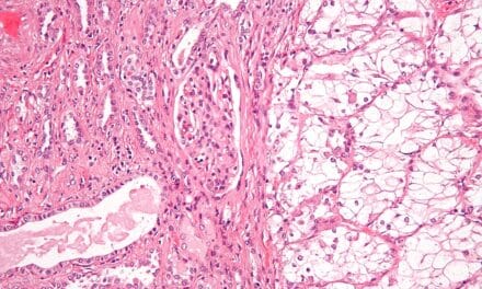
Featured image: PARPi-FL imaging of a clitoral excision – invasion from a vulvar squamous cell carcinoma. Imaging with PARPi-FL was also carried out in one patient harboring a recurrent squamous cell carcinoma of the vulva invading the clitoris. (A) Patient biospecimen ex vivo. The dotted area (1) corresponds to the tumor and the surrounding area (2) corresponds to benign margins. Bottom row: Images acquired with the handheld confocal microscope demonstrated tumor cells with enlarged nuclei that were different in size and arranged in a very disorganized manner. Tissue depth 4 μm (tumor) and 6 μm (normal tissue). (B) H&E staining showed that the tumor was restricted to a small area in the tissue – represented by number 1. PARP1 IHC corroborated the confocal findings since no PARP1 expression was present in the stroma (2) whereas it was present in tumor (1). (C) Quantification of PARPi-FL uptake in tumor and normal tissue. Fluorescence signal was significantly (p < 0.0001) higher in the tumor area when compared to the normal surrounding tissue.