 The last two years have been good ones for nuclear medicine, as the modality looks to rebound from the doldrums of the early 1990s.
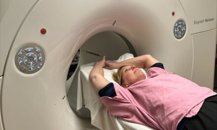
Left: GE Medical?s functional anatomical mapping combines the Millennium system with X-ray.
In 1997, the modality achieved a 10 percent growth, while 1998 produced an expansion of 9 percent.
Additional reimbursement approvals from the Health Care Finance Administration for several oncology-related positron emission tomography (PET) indications helped make PET a more appealing option for healthcare providers.
In addition, advanced techniques, new applications and the increasing availability of the radiotracer fluorodeoxyglucose (FDG) for dedicated PET scans also has propelled nuclear medicine?s resurgence.
?The outlook is that there may be broad [reimbursement] coverage for FDG over the next year,? noted Randy Whitehead, Siemens? vice president of nuclear sales and marketing.
Frost & Sullivan (Mountain View, Calif.) estimated the total nuclear medicine market at $694.3 million in 1998, but that level remains just short of the $700 million-plus levels in the early 1990s. Analysts predict the market will grow to $800 million this year and to $930 million by 2004.
At Siemens Medical Systems Inc. (Iselin, N.J.), much of the nuclear medicine talk was about the company?s E.Cam flagship product. Since its introduction four years ago and collaboration with TAMS two years ago, Siemens has introduced six E.Cam models ? the variable-angle, the fixed 180, fixed 90 for cardiac, E.Cam+ coincidence version and two single-head models.
With the help of a worldwide installed base of some 800 E.Cams, Siemens asserts that it has recaptured the No. 1 nuclear medicine position with a 30 percent market share.
Siemens also claims a nearly 70 percent worldwide market share in PET with its joint venture company, CTI Pet Systems Inc. (Knoxville, Tenn.).
ADAC Laboratories Inc. (Milpitas, Calif.) introduced its new SkyLight, a gantry-free nuclear medicine gamma camera. The works-in-progress allows the gamma detectors to be mounted directly into a room structure, removing limitations associated with the floor-based mechanical gantries of existing gamma cameras.
ADAC is calling the technology FreeDimensional imaging which allows the detectors to move in any direction together or independently. The system moves to accommodate a patient?s position rather than having the patient positioned to fit the camera.
ADAC said the concept will improve productivity and allow for new nuclear procedures, such as total body venograms, dual-detector total body spots, full brain SPECT and standing total body imaging. SkyLight?s openness and flexibility is designed for pediatrics, claustrophobic patients and lymphoscintigraphy.
SkyLight has been under developed off and on for the last two years. Work on the system started in earnest in the middle of this year in anticipation of its RSNA debut.
Mohamed Elmandjra, ADAC?s vice president of marketing, estimated that SkyLight could reach the market in the next 12 to 18 months, depending on the results of clinical trials and FDA review. The company has yet to file with the FDA.
SMV America (Twinsburg, Ohio) turned some heads on the show floor with its works-in-progress Positrace dual-mode oncology system.
Positrace combines a dedicated PET camera optimized for whole body FDG imaging and a CT scanner supplied by Analogic Corp. (Peabody, Mass.). The PET scanner has an opening of 70 centimeters. The PET camera includes six detectors in a hexagonal configuration, while the CT offers a two- to four-second rotation speed and produces 10-milimeter slices.
?We anticipate the street price for the device to be $900,000, including the CT scanner,? said Lonnie Mixon, SMV?s vice president of worldwide marketing.
Positrace is designed for whole-body FDG scanning because of its extended coverage. The axial coverage is 50 centimeters for one scan and 78 centimeters in two scans.
The Positrace has not received FDA clearance. The first two clinical sites are expected to be in France in the third quarter. SMV plans for commercially availability before the end of the year.
 Marconi?s Irix in three-detector configuration
The combining of imaging modalities also is evident in Marconi Medical Systems Inc.?s (Highland Heights, Ohio) integrated oncology center. The configuration combines the company?s nuclear medicine, CT and MRI technologies to improve the detection, diagnosis and treatment of cancer.
The solution includes Marconi?s variable-geometry, triple-detector Irix nuclear gamma camera, the AcqSim 3D simulation system and AcqPlan, an advanced dose computation system to maximize radiation to diseased areas, while sparing normal tissue.
Marconi has three installations of the oncology suite. The oncology suite ranges in list price from $500,000 to $1.5 million, depending on options and features.
Marconi also has received FDA 510(k) clearance to market its Beacon S. Beacon P clearance was received in May. Beacon S corrects for non-uniform attenuation during SPECT (single photon emission computed tomography) studies. Beacon P corrects for non-uniform attenuation during PET (positron emission tomography) studies.
Both Beacon S and Beacon P are available on Marconi?s Axis and Irix nuclear cameras. The device uses a scanning point source of radioactive Barium 133 to create the transmission map. The result after correction is an enhanced representation of the emission data. Plans are to begin shipping Beacon S this month.
Marconi also featured its gPET3 triple-detector coincidence-imaging product. By using all three detectors, gPET3 improves PET performance and enhances geometric uniformity across the field-of-view for better lesion detectability at the periphery of the body.
Toshiba America Medical Systems (TAMS of Tustin, Calif.) exhibited a high-end solution for whole-body, emission tomography, coincidence and general imaging procedures.
The system consists of TAMS? E.CAM+ variable-angle gamma camera and its GMS-5500A/PI workstation. The dual-purpose E.CAM+ performs routine nuclear medicine and high-energy coincidence imaging. Based on the Sun UltraSparc CPU, Toshiba?s GMS-5500A/PI workstation is designed for rapid processing of nuclear medicine procedures for greater clinical utility and productivity.
Clinicians can use the same system for full-featured SPECT and coincidence imaging. The company?s new product features and clinical software applications include UltraSPECT, a new program that automatically processes myocardial SPECT studies, reducing the manual steps normally required for myocardium reconstruction defining the angles of the ventricular chambers.
TAMS? new Merged SPECT (3D) Processing feature is a SPECT whole-body technique developed using up to five SPECT passes over the body. The data set is processed into one axial whole-body 3D image, instead of the traditional whole-body scan in a planar format that provides a skeletal view showing hot or cold spots with 2D localization.
Functional anatomical mapping continues to draw much of the attention in GE Medical Systems? (GEMS of Waukesha, Wis.) nuclear medicine arena. The FDA-cleared technique combines nuclear medicine images of a patient?s metabolism with images of his or her anatomy. The procedure allows clinicians to combine the separate images into one to provide more detailed information to detect and locate disease in the body.
GEMS still targets June 2000 for market release for the as-yet unnamed product. GEMS also introduced a high-performance, cancer-detecting PET scanner, which demonstrates biological function in organ systems.
The GE mobile PET Advance features a wireless data transceiver that shares patient data without the need for traditional network connections. Patients can be scanned in a mobile unit and have their images and information immediately transferred to the hospital electronically using PET Direct.


