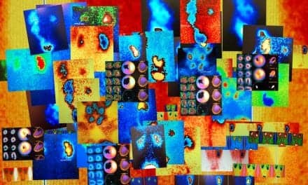
A study published in the journal Nature Nanotechnology has identified a new, noninvasive method for imaging the gastrointestinal (GI) tract. A multi-institutional research team has developed a nanoscale agent that relies on photoacoustic imaging and PET technology, possibly leading to improved diagnosis and treatment of gut disorders.
The research team included Weibo Cai, a specialist in PET imaging at the University of Wisconsin-Madison; Jonathan Lovell of the State University of New York at Buffalo; and Chulhong Kim at Pohang University of Science and Technology in South Korea. Lovell and Kim both specialize in photoacoustic imaging, which uses ultrasound and light-based imaging to create scans.
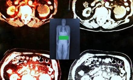
Intestinal disorders such as small bowel bacterial overgrowth, irritable bowel syndrome, and inflammatory bowel disease can lead to significant side effects in patients with diabetes and Parkinson’s. Current intestinal imaging methods rely on patients consuming a chalky liquid known as barium while technicians use x-rays and ultrasound to view the intestine. However, drawbacks to this method include potential radiation exposure and limited accessibility. In the new procedure, patients drink a liquid containing nanoparticles and bright dyes to illuminate and study the intestine in real time.
Researchers have completed studies with mice and anticipate moving into human trials soon. “We could potentially induce a paradigm shift that allows for much more routine examination of the intestine function,” Lovell said. “That would really benefit overall health.”
For more information, visit Nature Nanotechnology.