 GE Medical Systems’ Discovery LS PET/CT system with clinical images. Clinical images from top left: PET image, CT image and PET/CT combined image.
Location has always been a focus in the real estate market. A property’s appearance, although important, is just part of the consideration. When it comes to imaging information, location also plays a key role: Knowing the metabolic characteristics of a tumor just isn’t enough; identifying the location, the physiologic information, by incorporating CT with PET or SPECT is a growing area with expanding appeal.
Interest in hybrid applications has resided in the area of oncology, but are cardiology and neurology preparing to move into the neighborhood as well? Industry watchers have various opinions. However, all recognize one fact: Hybrid and multimodality imaging will continue to play a key role in diagnosis and management of disease.
A new analysis from Frost & Sullivan (San Jose, Calif.) reveals that the U.S. PET and PET-CT markets generated revenues totaling $481.23 million in 2002. The report estimates total market revenues could reach over $1 billion dollars in the next three years.
“The emergence of PET-CT has usurped sales of stand-alone PET but has added robustness to the total market to keep it thriving,” says Frost & Sullivan Industry Analyst Manager Monali Patel. “Increasing evidence indicating the superior ability of PET-CT to detect a number of cancers and be used in conjunction with radiation therapy planning is adding to the excitement in the marketplace.”
Predictably, challenge accompanies technological change. Combining PET and CT images makes the scanner operation complex, and the shortage of cross-trained staff in nuclear medicine and radiology at facilities poses another challenge to adoption, according to the Frost & Sullivan report.
Turf issues will continue to complicate matters. Who should control the devices remains an issue. Should it be the radiology or nuclear medicine group, or a shared responsibility? But the power of multimodality imaging in general and the potential of research tools, including optical imaging, conjure more promise than opposition.
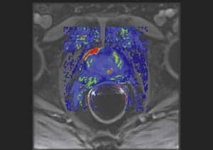
 Siemens’ biograph Sensation 16 PET/CT system. Clinical images show an increased activity in the right anterior wall in a 46-year-old male with a history of bladder cancer.
“Data show and research will show that CT alone will give the position about 60 percent diagnostic accuracy,” says Steve Bolze, general manager Global Functional and Molecular Imaging for GE Medical Systems (GEMS of Waukesha,Wis.). “If you use PET, it goes to 90 percent diagnostic accuracy. PET-CT will take it to 98 percent diagnostic accuracy in the case of lesion detection. So what’s really going on in terms of the advancements in PET is the integrated or hybrid PET-CT, because you get the highest diagnostic confidence and accuracy [for] lesion detection.”
GEMS introduced its Discovery ST, its first fully integrated PET-CT for cancer, at last year’s RSNA. The Discovery ST provides 2D and 3D imaging and has been optimized for larger patients and to facilitate radiation treatment planning. The Discovery LS, the initial GEMS PET-CT technology, provides 2D imaging capability. More than 100 PET-CT systems are installed worldwide.
GEMS also offers SPECT-CT technology in its Discovery VH. The system includes a high-end nuclear camera for SPECT imaging and the Hawkeye CT component for attenuation correction.
Contrasting approaches
Vanderbilt University Medical Center (Nashville, Tenn.) does approximately 30 to 40 PET-CT scans per month, with the number growing daily. It uses the Discovery LS from GEMS. The technology involves a multislice CT system, which is configured back to back with a PET scanner. The multislice scanner is used to transmit images for attenuation correction of the PET scan, providing anatomical localization of the PET images.
“That does two things: You’re getting attenuation correction of a PET image and at the same time you can reconstruct the CT data to allow you to take your PET images, which can then be fused with your noncontrast-enhanced CT scan to give you a more specific anatomic localization,” says Martin Sandler, M.D., chairman of radiology and professor of radiology and radiological sciences at Vanderbilt University Medical Center. “And that is really in my opinion going to become the standard way that PET studies are going to be read in the future.”
 Siemens’ E-CAT Excell
Sandler says there is a lot of Gestalt to reading images: Is something an artifact? Is something in the valve or in the lymph nodes? Where exactly is it? “The accuracy of reading PET, as good a modality as it is, is somewhat limited in the hands of the inexperienced and … the experienced,” Sandler says. “There’s a certain amount of ‘I’ve seen a thousand cases and this is what I think it is.’ But when you can fuse it to a CT image, it improves the accuracy tremendously, even in a noncontrast-enhanced image.”
Some people are doing contrast enhancement at the time of their PET images. In other cases, a diagnostic study is done after they’ve done their images by giving intravenous contrast. “I think that is the Cadillac of oncologic imaging,” Sandler says. “It allows you to have a patient done at the same time on the same machine that gives you the most accurate anatomical fusion.”
Moving forward
Radiation therapy continues to garner increasing attention. “Now they have intensity modulated radiation therapy that they can very accurately target the radiation therapy beam, and more and more of the advanced radiotherapy treatment planning centers are using a combination of PET-CT to treat the tumors in patients that have good fluoro-deoxyglucose uptake,” Sandler says.
The anatomical region of the tumor does not always accurately represent the area of active tumor growth and by doing a combination of metabolic imaging and anatomical imaging, you can more precisely localize the area of tissue that is active tumor vs. debris and surround edema and swelling at the active tumor center.
 Gemini PET-CT scanner from Philips
“Like any sort of technology that first gets delivered to the marketplace, we’re seeing added sophistication year by year,” Sandler says. “The basic concept is not going to change, but I think CT and the PET scanner are getting more sophisticated, and now there are systems that are being designed that will do a total body scan in 10 minutes. We’ve gone from 45 minutes to doing a total body scan with attenuation correction to the 10- to 15-minute range. That’s pretty impressive.”
With multislice CT, a whole body scan can be done in 30 or 40 seconds. Sandler sees the speed of acquisition as a challenge for the PET-CT system. “I think the challenges of taking a spiral CT scan, which is done in seconds, and fusing it with a modality that is done over 10 minutes collection results in some degree of image fusion which is not totally aligned,” Sandler says.
A multi-center trial continues on the use of CT- SPECT to provide attenuation correction for cardiac imaging. Sandler says preliminary data show that it is “fairly accurate.”
Currently, Sandler uses CT-SPECT for imaging a variety of oncological tumors and infections. “The quality of the CT scan right now is really nondiagnositc and just more to give anatomical landmarks,” Sandler says. “There’s certainly very limited diagnostic interpretation, and differentiation in many instances between different tissue masses is not quite possible, so I think the improvement in the sort of gamma camera technology quality of images [is needed], but I think for CT-SPECT to be a viable product, the quality of the CT scan is going to have to improve.”
CT-SPECT brings greater value to the utilization of SPECT on its own. The ability to localize abnormal focus of activity to a specific organ as opposed to an area of the abdomen, for example, positively impacts surgery. For patients with thyroid cancer, the surgeon makes a small incision and removes the tumor, avoiding the need to explore the neck. Patients can return home the same day.
The recent focus on cardiac attenuation, developing various algorithms and bringing the technology to multi-center trial has been an important step. “Trying to make a viable product that will be reimbursed is a very important step,” Sandler says. “I think one of the problems with both CT-PET and CT-SPECT is that unless one does a diagnostic-quality study with contrast, etc., it’s adding tremendous value. At some point, the need for a CPT code for either attenuation and/or anatomical fusion is going to be necessary to move or sustain utilization of the product, because it is providing greater patient diagnostic information. It’s a reality that needs to be filled.”
Sanjiv Sam Gambhir, M.D., Ph.D., director of Crump Institute for Molecular Imaging (Los Angeles) and professor in the Department of Molecular and Medical Pharmacology, Department of Biomathematics at University of California Los Angeles (UCLA) School of Medicine uses the CTI Molecular Imaging Inc. (Knoxville, Tenn.) and Siemens Medical Systems (Malvern, Pa.) biograph PET-CT system.
Gambhir’s clinical work in body oncology includes lung, breast, colorectal, head and neck cancers, melanoma and lymphoma. “For all these application areas, the key is for years we read the FDG PET image alone and then sometimes had correlative CT scans on the patient done either in an outside hospital or here on a separate CT scanner,” Gambhir says. “It’s very different to register the information because the information is taken on each separate scanner. There were no good software tools for merging the information for the whole body.”
Using small animal models, Gambhir and his colleagues study multimodality imaging to better register the mouse heart and blood flow to the heart, as well as implantation of stem cells to the heart. “Multimodality MR-PET, which is also being developed, might be useful for clinical applications in cardiovascular areas, because then you could image the heart as it beats along with the information of blood flow that the PET part can give you,” Gambhir says. “But PET-CT as opposed to PET-MRI for clinical cardiovascular, it’s not clear to me how that would be useful at this stage.”
As for the future of PET-CT scanners, Gambhir anticipates that almost all PET scanners will be replaced by PET-CT. “There will be no [single]-modality PET other than at a few research centers that might need very high resolution PET,” Gambhir says. “We think for clinical reasons and applications, essentially every PET scanner that will be sold will be a PET-CT. There is no reason to use just PET alone when you can have both anatomical and functional information. We think the exponential growth is just beginning.”
Several factors will drive PET-CT, including better software tools to analyze both CT and PET and new tracers. “It’s difficult to go back and forth between CT information on these fused images,” Gambhir says. “There’s a lot of software development that needs to take place. We also think a lot of new tracers that are more specific so they only home in on specific molecular targets [will play a role]. These will continue to accelerate and further fuel the need for PET-CT, because if you have a tracer that only goes to certain sites and that’s all that lights up, the only way to know where it lights up is by having the CT information superimposed on it. We think the new tracer development will drive PET-CT further.” Minibodies is one example.
Whether to administer contrast orally or through an IV during the PET-CT scan remains an issue. “Those are important because they will make the CT quality better and the CT portion more interpretable on the PET-CT scan,” Gambhir says.
Most places doing PET-CT are not yet incorporating contrast for CT. It is technically more difficult. There’s possibly interference of the PET image from the contrast as well.
“It is an important area that has to be resolved,” Gambhir says. “It’s just that it hasn’t been appropriately tackled either by the vendors or by the people doing the studies.”
Multimodality imaging will continue its move into neurology. “There it’s PET-MRI because the brain anatomy is much better visualized for most processes with MRI instead of CT and PET,” Gambhir says. PET can distinguish radiation necrosis from recurrence of tumor, for example. MRI cannot.
“Alzheimer’s disease, which PET is very good at [imaging] could be [viewed] by correlating PET with MRI, but there’s currently not a clinical MRI-PET system,” Gambhir says.
Better ways
Optical or molecular imaging uses light emitted from the patient and uses no radioactive materials. “The optical has some synergy when used, for example, with PET, so we are in the process of looking at designing systems that will merge optical and PET detectors,” Gambhir says. “Currently the prototypes are being built for small animals, but the hope is to translate that into some environment where optical-PET will be useful in other arenas as well.”
Animal prototypes for optical PET are approximately two years away. Clinical systems are about seven to eight years out. Elsewhere, work continues on merging optical with CT. Gambhir says it’s not out of the realm of possibility that optical will also merge with MRI.
The early expectation of multimodality imaging was that PET-CT was going to provide perfect registration. But reality is beginning to set in, according to Horace Hines, Ph.D., chief technical officer for Philips Medical Systems Nuclear Medicine (Bothell, Wash.).
“Even though they’re on the same instrument, the patient still breathes; he or she still has peristaltic motion in the gut, and there are muscular contractions that go on that you’re not even aware of, and that causes motion in the lower abdomen,” Hines says. “These processes happen in seconds to minutes, and when you’re talking about studies that are separated from each other by as much as 10 to 20 minutes, you get motion between the anatomical imaging and the physiologic imaging.” That points to the greater need for image registration software to improve alignment.
In addition, the perfect registration isn’t true because of the physiologic motion and the perfect attenuation correction isn’t true because of the lack of registration and the lack of correct mapping of the attenuation caused by contrast material. “That’s the area where we are making advances,” Hines says. “The advances are improved registration and confidence in the physician interpretation by virtue of having the CT and the physiologic data, because [prior to the multimodality approach] often the physician interpreting the PET study didn’t have the CT study.”
The industry overall is working to make instruments more efficient in terms of collecting more counts in a shorter time. Image quality and improvement of registration software also remain in the forefront.
Philips’ answer to the registration challenge is its Syntegra registration and fusion software. The software allows the radiologist and radiation therapist to simultaneously view the image, consulting by phone, and ensure the correct place is being irradiated or point out an area that the therapist might not have thought to radiate but should.
When it comes to cardiology, interest continues in the 16-slice CT to get combined perfusion and anatomical information using CT angiography for the coronary arteries. The need for registration in the heart is really not as great as it is in oncology, and I don’t think people have spent a lot of time thinking about that yet in analyzing what machine they should get and how costly it is, Hines says.”
Siemens is looking for its biograph Sensation 16 to move the cardiac market forward, according to Jonathan Frey, manager for PET marketing and sales support. The system uses lutetium oxyorthosilicate (LSO), the most recent in crystal technologies for PET. Siemens introduced LSO in 2000 on its ECAT Excell, a dedicated PET scanner.
“It shows distinct benefits in cardiology, allowing you to do a complete cardiology workup, both the CT angiography for occlusion detection, as well as viability and perfusion on the PET side, all within about 20 minutes,” Frey says. “For a typical oncology exam, you would also be able to do the entire exam, both PET and CT, in one go in roughly less than 20 minutes.”


