Two New Software Offerings from GE
GE Healthcare, Waukesha, Wis, released two new software packages for advanced analysis of cardiovascular magnetic resonance (MR) images: CardiacVX and MR VesselIQ Xpress. The company’s CardiacVX software delivers fast, intuitive, semi-automatic analysis of key cardiac parameters, allowing the user to create a comprehensive cardiac patient report within a streamlined clinical workflow. MR VesselIQ Xpress, based on GE Healthcare’s CT VesselIQ Xpress platform, is an optimized image analysis package for MR angiographic data. It analyzes selected vessels for stenosis, directional tortuosity, and other vascular anomalies. MR VesselIQ Xpress provides advanced tools such as automatic vessel tracking with centerline display of any vessel, quick 3D visualization, and fast access to vessel cross-section and profile images.
ViZion + DR Panels from Viztek
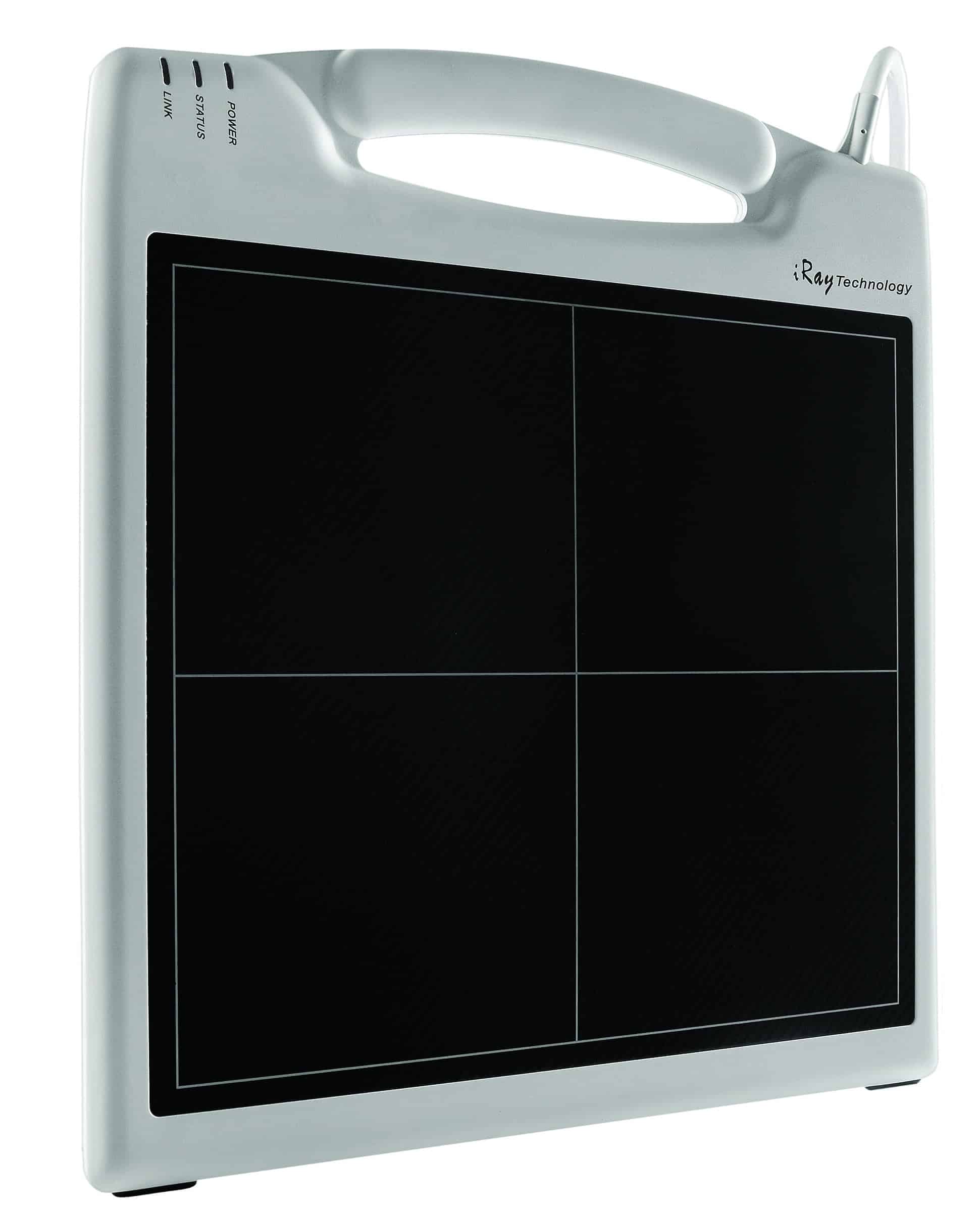
Viztek, Raleigh, NC, the advanced digital imaging solution provider, has 510(k) clearance for the ViZion + DR line of panels. This new line of products includes multiple panels in tethered, wireless, and fixed configurations offering high quality images at quick speeds. The panels require no generator interface. The ViZion + DR panel product line has a versatile range of applications, including new x-ray rooms, retrofit rooms, and mobile implementations. The fixed panels will be available in Viztek’s Straight Arm and U-Arm configurations.
FDR Go from FujiFilm

FujiFilm Medical Systems USA Inc, Stamford, Conn, reintroduced the FDR Go at RSNA 2012, representing the latest evolution of its digital portable portfolio. Fujifilm’s new DR models have evolved to a next-generation level with advanced Fujifilm DR features such as irradiated side sampling (ISS) and dynamic visualization (DV). ISS senses x-rays at the top detector layer, minimizing light spread (blur) and capturing sharper signals. DV clarifies images for higher visibility detail and enables wider range of window and leveling throughout the entire image. The FDR Go is designed for image preview in as fast as 1 to 2 seconds. Clinicians can then send images wirelessly to the hospital network without leaving the patient bedside. Features designed to simplify workflow include: SpeedLink, which automatically maps generator techniques, tube head inching, and spare battery charging.
Postprocessing and Structured Reporting from RamSoft, Digisonics

RamSoft, Toronto, has partnered with Digisonics to offer integrated, vendor-neutral, cardiac postprocessing and structured reporting for all cardiovascular modalities. Digisonics was recently named Best in KLAS for the cardiology market segment for the fifth consecutive year. RamSoft’s PowerServer RIS, PACS, and teleradiology solutions transfer the relevant patient studies via DICOM to the Digisonics workstation for high performance image analysis and professional reporting, which can be completed in minutes. This integration maximizes efficiency and productivity by automating the complete workflow for cardiac specialists.
Clinical Viewer from DICOM Grid
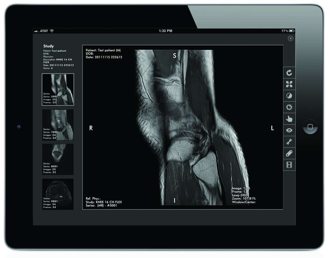
DICOM Grid, Phoenix, Ariz, a global provider of cloud-based medical image exchange solutions, recently launched their next generation clinical viewer. This latest release enables the easy access and viewing of complex medical imaging data and associated reporting from any browser-based device. Mobile viewing enables doctors to streamline collaboration with patients. The viewer comes equipped with a variety of interactive tools used for measuring, annotating, and manipulating images. It can even be optimized for low bandwidth environments.
Transthoracic Echocardiography Simulator from HeartWorks
In echocardiography, images of the functioning heart are obtained through a transthoracic procedure, a handheld probe placed over the chest, or a transesophageal procedure that creates close-up images of the heart via a probe guided through the mouth into the esophagus. A new simulator from Heartworks, London, could make training for the procedure more effective. The simulator, which uses Ascension Technology sensors, utilizes a lifelike manikin torso, visualization software, and an ultrasound probe to provide training in both procedures. Using the ultrasound probes, trainees gain practice obtaining varied views and interpreting 2D ultrasound images while working in the context of the 3D structures of a beating heart. The simulated 2D and 3D images are displayed side-by-side on a computer screen.