|
According to 2007 research by the Centers for Disease Control and Prevention (CDC), the percentage of women aged 40 and older in the United States who had a mammogram in the last 2 years varies from state to state. The states with the lowest breast cancer screening percentages (67.3% to 72.8%) include Arkansas, Colorado, Idaho, Indiana, Mississippi, Missouri, Montana, Nevada, New Mexico, Oklahoma, Texas, Utah, and Wyoming. The states with the highest screening percentages (79.2% to 84.8%) include Connecticut, Delaware, the District of Columbia, Maine, Maryland, Massachusetts, Michigan, Minnesota, New York, North Carolina, Rhode Island, Tennessee, and Vermont.
While physicians and advocates of women’s health would like to see those numbers increase even more for the sake of better patient outcomes, there’s one thing industry professionals can agree on: Mammography reading is not for the faint of heart.
The sheer volume of mammographies that are performed—and need to be read—makes for a demanding workload. For radiologists across the country, it’s a quandary. You might be based in a bustling metropolis where sophisticated urbanites are focused on their health, and so population exponentially increases your reading load. Or, you may be based in a remote, rural area where the shortage of breast specialists puts your hospital or imaging center under additional strain because you simply don’t have the human resources.
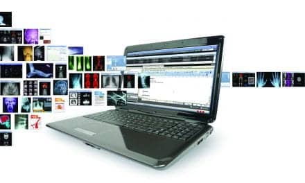
A host of recently launched remote reading solutions is the latest way to keep pace with mammography. Here are just three case histories that illustrate what today’s innovative technology can do for your patients, your practice, your breast center, or your community hospital.
AIMING FOR FLEXIBILITY
Let’s say you run a hospital and you do mammography on your main campus, but your organization also performs mammography exams at an affiliated hospital across town and at a breast center. Your mammography readers are housed at different places and have the availability to read at different times. So the challenge is making sure you can efficiently transfer full-field digital mammography (FFDM) images to wherever they need to go—at any given moment.
A tall order? Yes, but doable, says Phil Ames, director of radiology, St. John Medical Center in Tulsa, Okla. Ames said the key is to aim for flexibility. “The real problem is that most digital mammo workstations from manufacturers are just that. We needed something that worked for mammography, but also CT scans and so on,” Ames said.
That’s why St. John is now implementing multimodality breast imaging workstations from Carestream Health. The goal is to enhance efficiency since radiologists need to read diagnostic and screening mammography exams as well as general radiology exams on the same workstation.
For Ames, the first hurdle was speed. Thanks to a metropolitan Ethernet connection, “it now takes just 5 minutes for a mammography to get across town to here,” Ames said. But previous patient data is critical, too. “With our Carestream PACS we can pull up a patient’s entire folder—priors, biopsies, whatever it is that the physician needs in order to make a clinical comparison.”
But the workstations—”the front end of the PACS system,” as Ames puts it—were truly critical to the efficient reading of mammographies. “The challenge was that a lot of the manufacturer workstations are set up to handle full-field digital mammography. In other words, they have to be labeled as FFDM,” Ames said. “We needed a workstation that can recognize or understand a digitized prior. Carestream’s multimodality workstation solves that problem.”
St. John went digital less than 2 years ago. Today, all screening mammograms are captured on seven FFDM systems at the health system’s multiple locations. “Since going digital—and with our Carestream PACS and multimodality workstations—we’ve gone from performing approximately 21,000 screening exams a year to nearly 29,000,” Ames said.
The multimodality workstations solved a multitude of problems for clinicians at St. John. According to Ames, all too often, technology is inefficient. “Sometimes there is just a lack of information for the radiologists,” Ames said. “For example, you may not have the right RIS to pull up a patient’s history, and then the radiologist has to wait for paperwork.” But the Carestream PACS and multimodality workstation, Ames says, can access both HL7 and DICOM information—so the radiologist can find everything he or she needs to read and diagnose.
TRY HOLDING A CANDLE TO THIS
Based in Danbury, Conn, Candlewood Center for Women’s Health is dedicated to providing a full range of women’s health services. For many years, the center offered analog mammography. Recently, Candlewood upgraded to FFDM acquisition. But without an on-site radiologist, the group continued to face the big challenge of delivering timely exam reading and reporting.
 |
| ©Bruce Robinson |
With the analog system, Candlewood’s process was laborious. The group printed films and relied on couriers to transport them to and from offices of its off-site radiology reading group, Northeast Radiology in Brewster, NY. After the digital transition, the practice planned to send images electronically for faster turnaround but did not need a full PACS installation.
That’s when Candlewood turned to CoActiv for a workflow and infrastructure installation solution. According to Amy Kohn, practice manager for Northeast Radiology, Candlewood’s goal was ambitious. “The system had to enable rapid access to large digital mammography files for Northeast’s multiple offices in both New York and Connecticut.”
CoActiv customized a system that allowed files to be sent seamlessly to Northeast’s offices, wherever needed. Kohn notes that the radiology practice’s workflow is complex, with multiple imaging sites and sub-specialty radiologists. CoActiv’s teleradiology system easily provided flexible image access to ensure prompt exam turnaround.
A key challenge concerned HIPAA. Its regulations prohibit mammography files from being compressed at any time in the life of the study. CoActiv addressed this challenge and crafted a solution that provided rapid transmission of image files in full fidelity.
The new process: Digital files are sent from Candlewood’s Hologic modality to an innovative on-site CoActiv Acquire & Forward Server. This server provides automatic and secure data communications over the Internet directly to a CoActiv Exam-PACS server at Northeast’s main location. Following this, the exams automatically appear on Northeast’s radiology worklists.
Simultaneously, all images are saved to CoActiv’s Exam-Vault quad-redundant archiving in four distinct instances, for fail-proof disaster recovery and business continuity. Once exams are read, reports are immediately available electronically to Candlewood and are also saved with the images to the CoActiv archiving system.
Kohn says image communication is seamless and fast, and information has flowed flawlessly since the system was installed. Candlewood and Northeast Radiology agree—no other vendor could hold a candle to their CoActiv solution.
BRINGING IT HOME
In 1991, Elizabeth Pusey, MD, founded Women’s Medical Imaging in Newport Beach, Calif, to provide advanced digital mammography services and other women’s health services, such as ultrasound, to her community. In 2006, the practice adopted a Siemens Mammomat NovationDR full-field digital mammography system to accommodate referring physicians needing high-quality breast imaging and critical diagnosis in order to develop comprehensive treatment plans for their patients.
Once Pusey had a digital mammography system up and running at her practice, she sought a solution that would enable her to remotely read imaging studies. She believed that if “telemammography” could be a reality for her practice, it would afford her the flexibility and convenience of working from home.
Pusey began her search for the right solution. She considered a number of PACS solutions but was deterred by the high cost and complexity. Next, the practice tried to maintain a CD-based digital imaging archival and retrieval system, but this approach proved to be inefficient and called for significant manual operation. In addition, it did not provide the practice with immediate access to prior studies and lacked the scalability to keep pace with the workload.
Ultimately, the solution for Pusey and Women’s Medical Imaging came in the form of the Candelis ImageGrid PACS Appliance system. Here is what Hossein Pourmand, vice president of business development, Candelis Inc, had to say about getting Pusey’s organization up and running: “The 1.5-terabyte raw capacity system, which can accommodate approximately 45,000 digital mammography studies using a 4:1 lossless compression, was installed in just 4 hours with no interruptions to daily operations.”
The Siemens digital mammography and the ultrasound systems send images directly to ImageGrid, and studies are immediately available for diagnostic viewing. What’s more, the ImageGrid is DICOM-compatible, which meant that incorporating with the existing Siemens MammoReport workstation was seamless.
But what about reading from home? Pusey’s practice averages more than 20 studies per day, and the number is expected to increase.
Digital mammography studies can range between 80MB and 100MB in size, requiring a system that can accommodate the transfer of large amounts of data. ImageGrid Mammography Web Viewer gets the job done because it is a thin-client application with automated rule-based, prefetching capabilities that significantly improve the data-transfer speed and access to prior studies.
In April 2008, Pusey took the leap. The work-from-home setup was quite easy. “With her existing ImageGrid system, the addition of the ImageGrid Mammography Web Viewer required a simple software installation, proper configuration, and calibration of displays at her home office,” Pourmand said. Now, Pusey can provide timely reports to her patients and referring physicians from the comfort of her own home.
The ImageGrid Mammography Web Viewer is a Web-enabled application residing on the ImageGrid PACS appliance. It gives physicians the flexibility and convenience of viewing mammography studies from any DICOM workstation on a Local Area Network, Wide Area Network, or remote workstation via a Virtual Private Network.
“The complete solution addresses critical archiving, image management, workflow, visualization, reporting, and report-distribution needs on one single hardware platform,” Pourmand said. What’s more, the solution addresses the needs of imaging centers as well as radiology/women’s imaging divisions within large hospitals. And for Pusey, well, Candelis brought home precisely the solution she needed.
—Marianne Matthews
Chill Out!
For some women, a mammogram can be painful, even traumatic. In fact, it can be a pretty chilling exam. Now, a new product is helping to make mammography a bit more comfortable. “We’ve received comments from our patients on the decrease in the coldness of the machine. … They are easy to use, economical, and pretty—all at the same time,” said Susan Bennett, RT, RM, ARRT, Colquitt Regional Medical Center, Moultrie, Ga. “We love our Bella Blankets, and so do our patients.”
The Beekley Corp, based in Bristol, Conn, recently introduced Bella Blankets Protective Coverlets for Mammography. Designed to remove the chill from the bucky/receptor plate, the company says Bella Blankets Protective Coverlets help patients feel more at ease with the procedure and improve patient-satisfaction levels by drastically minimizing complaints that the bucky is cold.
“Patients do not complain about the sharp feeling of the cold bucky. This leads to patient satisfaction, which is very important to me as a technologist,” said Anita Blalack, mammography technologist, Marble Falls Imaging Center, Marble Falls, Tex. “The product also helps with the perspiring patient to immobilize the breast as well as calming the nervous patient.”
IDEAL FOR SPECIAL NEEDS
Comfort is one issue, but clinical benefits are another. According to the Beekley Corp, Bella Blankets’ fabric-like material may also help with positioning by immobilizing small breasts or those prone to perspiration. In addition, the product provides added protection for patients with thin or delicate skin, and those who have cuts in the inframammary fold without affecting image quality.
Single use and easily disposable, Bella Blankets Protective Coverlets are FDA approved and compatible with both digital and analog units. The product is available in two sizes and an elegant floral design. To learn more, and for complimentary samples, call (800) 233-5539 or visit www.beekley.com.
A mammogram may not be a manicure, but a woman should feel anxiety-free. Thanks to Bella Blankets, women across America can now chill out during their next exam.
—M. Matthews