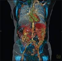
More than a century ago, physicians began using radiation as a tool to treat malignancies when x-ray was first used to treat a breast cancer patient. Since that time, the challenges have remained fundamentally the same—how to deliver the optimum dose to the tumor without hitting the sensitive organs and tissues surrounding it.
 Inside a medical linear accelerator from Varian Medical Systems. The radiation beam passes through and is shaped by a multileaf collimator so that it conforms to the contours of the tumor.
Improvements in radiation therapy in the last half of the 20th century have meant better treatment outcomes with fewer side effects for the patient. The increasing use of linear accelerators in the 1960s meant that physicians could turn the x-ray beam on and off during treatment, allowing greater control over targeting. Three- dimensional treatment planning, which gained ground in the 1980s, allowed for still better targeting of tumors while attempting to avoid surrounding tissues and organs.
All of this has paved the way for intensity modulated radiation therapy (IMRT), says Karen Marks, manager of the radiation oncology center at Baptist Memorial Health Care in Memphis, Tenn. She pronounces IMRT to be a technique still “in its infancy” but one that is already used in many types of cases—and is steadily growing.
 |
 |
 |
| Bottom Row, Far Right: Prostate: IMRT plan surface dose map: A plan for treating prostate cancer with IMRT concentrates the radiation dose in the tumor (red) with minimal exposure (yellow, green, and blue) to the nearby bladder and rectum. Bottom Row, Center: An IMRT treatment plan for treating a sinus tumor. The red area shows where the radiation dose will be concentrated. The eyes and spine, depicted in green and blue, will be avoided. Bottom Row, Left: A treatment plan for treating lung cancer. Radiation beams are delivered from different angles, and converge on the tumor, seen here enveloped in a “dose cloud.” | ||
What is IMRT?
IMRT is a technique made possible by a combination of computer and linear accelerator technology. The use of a multileaf collimator on the linear accelerator means that a number of leaves move in and out of the radiation beam in a customized pattern, shaping the beam to better allow it to enter the body and hit the tumor without hitting surrounding areas. The multileaf collimator delivers a carefully shaped beam as the beam gun moves around the body, hitting the tumor from every angle.
Some IMRT equipment makes use of other techniques to further refine control over the beam and allow for even more complex intensity maps. Scott Johnson, product manager for Varian Medical Systems in Palo Alto, Calif, explains that the Varian products make use of an “electrified screen at the front of the wave gun” that allows for rapid pausing and resumption of beam delivery—up to 20 stops and starts per second. Thanks to this screen and the fine control over the collimator leaves, “we can deliver a much more complicated beam,” Johnson says.
The importance of control of the radiation beam cannot be overstated, and it is the major difference between IMRT and radiation therapy techniques that have come before. “We [could] always cover the target,” says Johnson, explaining that all forms of radiation therapy can deliver the desired dose to the desired spot. It is the ability to “sculpt the dose away from sensitive structures” that is the hallmark of IMRT, Johnson says.
IMRT also makes use of a technique known as “inverse planning.” In traditional radiation therapy, a dose is picked for the beam gun to deliver, with the expectation that the correct dose will reach the target. With IMRT, the treatment planning physician chooses the dose most appropriate for the tumor—and the dose acceptable for any of the variety of surrounding tissues—and lets the computer work backward to decide the radiation delivered at every point in the treatment. Without the use of computers, this would be an impossibly cumbersome task.
Still, to achieve the maximum effectiveness, the planning can be an extremely complex process. The planning physician will go through each of the initial CT slices—often, 100 or more slices—to mark tumor sites to receive full radiation doses, tissues that can tolerate a moderate dose, and sensitive tissues and organs that can tolerate only a minimal dose or no dose at all. This process can be quite time-consuming, taking several hours and a great deal of experience. “[IMRT is] very expensive and time-consuming to plan,” says Marks, estimating that it may take an expert medical physicist some 16 hours over a week to 10 days to plan the treatment. But this complex planning can yield important benefits. By sparing surrounding tissues from radiation, IMRT can save patients from side effects ranging from visible skin burns and chronic dry mouth to loss of bladder and bowel control, to blindness.
State of the Art
IMRT is a technique that has the potential for more widespread adoption and a wider range of uses. This is the conclusion of a paper in a 2003 issue of the journal Cancer written by Loren Mell, MD, and John Roeske, PhD, both of the department of radiation and cellular oncology at the University of Chicago, with Arno Mundt, MD, of the department of radiation oncology at the University of Illinois at Chicago.
In a survey of randomly selected radiation oncologists, just a third reported using IMRT. This number was growing rapidly, however, as nearly 80% reported adopting IMRT since 2000. Academic radiation oncologists were more likely than those in private practice to use IMRT, partially because they may be using IMRT for research in addition to improving treatment to patients. The most common types of cases in which IMRT was used were head and neck cancers and genitourinary tumors, although other physicians were also treating central nervous system, lung, and breast tumors, as well as pediatric tumors, lymphomas, and sarcomas, with IMRT.
The study found that the most common barriers to using IMRT were lack of equipment and insufficient staff. Because of lack of equipment, some radiation oncologists do not have the opportunity to become familiar with IMRT, pointing to a need for more training. This training could, in turn, reap economic benefits for physicians’ practices, as patients increasingly seek out centers that offer IMRT. Ultimately, most of the IMRT users in the study planned to increase their use of the technique in the future, and nearly all nonusers expressed plans to begin using the technique.
 NOMOS’ radiosurgical package, STAT Rs, includes AutoCrane, an automated method for accurately indexing the position of the couch/patient during a radiation therapy treatment.
Who Will Benefit?
For some patients, IMRT is not appropriate. This includes cases such as Hodgkin’s disease and rectal cancers, both types of cancer where radiating the surrounding area will help to eradicate cancer that has metastasized, says Todd Barnett, director of radiation oncology for Swedish Cancer Institute in Seattle, Wash. “About 15% to 20% of patients will be candidates for IMRT,” adds Marks.
However, when the physician needs to steer the radiation dose around sensitive structures while delivering a high dose to the tumor, IMRT is the technique of choice. Barnett notes that he most frequently uses IMRT in head and neck cases, when it is necessary to spare the salivary glands from radiation to avoid chronic dry mouth and dental caries that can result from too much radiation. Barnett also frequently uses IMRT to treat prostate cancers, as the prostate is generally boomerang shaped and is nestled among highly sensitive areas such as the rectum.
And IMRT can bring with it an astounding degree of accuracy and flexibility, making it the choice to treat tumors that are unreachable any other way. “We can unwrap a tumor from around an optic nerve,” says John Manzetti, president and CEO of NOMOS Corporation in Cranberry Township, Pa, the first company to commercialize IMRT in 1994. This is an example of IMRT’s “submillimeter accuracy,” Manzetti says.
One of the newest types of treatment to benefit from IMRT is certain breast cancers. Michael LaCombe is a physician with Evanston Northwestern Healthcare (Ill), one of the few institutions in the United States to be using IMRT on the breast. Currently, IMRT is most appropriate for a fairly narrow range of breast cases: Evanston offers this treatment to postmenopausal women with tumors smaller than 2 cm (such as those found in ductal carcinoma in situ), lymph nodes negative for cancer, and clean surgical margins. These are the patients who often decide against follow-up radiation after surgery—LaCombe estimates that about a quarter of patients fitting this profile never receive radiation therapy, even though radiation decreases the chances of recurrence.
“Tumors tend to come back where the surgeon’s knife [has been],” says LaCombe. And IMRT allows the physician to better target the areas surrounding the former tumor site. “If the tumor is a peach pit, we’re targeting the peach,” he says. Better targeting has meant that Evanston Northwestern has reduced the course of follow-up radiation from 32 treatments to just 16 treatments over a 3-week period. Evanston Memorial has been employing IMRT for the breast for about a year and has had no episodes of recurrence and virtually no unpleasant side effects for the patients.
 CORVUS, the inverse treatment planning component of NOMOS’ premier IMRT offering, uses high-speed computer systems and a sophisticated optimization algorithm to allow the computer to determine the optimal method for delivering the physician-prescribed dose to the identified target while limiting dose to sensitive structures.
What Are the Drawbacks?
Carol Kornmehl is an attending radiation oncologist at The Valley Hospital in Ridgewood, NJ, and author of The Best News About Radiation Therapy (Academic Radiation Oncology Press, May 2003). Although she successfully uses IMRT in many of the most common cases—prostate, brain lesions, head and neck, and some breast cases—she notes some definite drawbacks to the technique.
“IMRT can create hot spots, or doses that are higher than the prescription dose, that are undesirable. For example, in standard radiation therapy, radiation oncologists generally keep the highest dose an area receives within 5%, or perhaps 10% when push comes to shove, of the prescription dose. IMRT hot spots can approach 30% to 40%,” she says. She also notes that “daily treatments with IMRT usually last for as long as 30 minutes, whereas conventional radiation therapy usually takes no more than 15 minutes. Therefore, patients who receive IMRT need to hold still on the treatment table for a relatively long time.”
Barnett also finds the lengthy planning procedure to be a drawback to IMRT. “[We] need to go through every CT slice and tell what needs radiation and how much,” he says, explaining that delineating the areas to avoid is as important as marking the areas to treat. For example, when treating a head and neck case, it is critical to mark the lips to be spared from radiation. Otherwise, the computer will believe that the lips are an appropriate path through which to pass radiation, and the patient can end up with uncomfortable burns.
 NOMOS’ BAT SXI utilizes an ultrasound system to image the target and associated organs immediately prior to treatment.
Currently, this marking is done manually by the physician planning the treatment, and only the axial plane is available to view the field for treatment. With the addition of more planes and more computer intervention, the process could be speeded considerably. “[We] need to be more automated and speed things up,” Barnett says.
James Adams, medical director of the radiation oncology center at Baptist Memorial Health Care Center, has also found that increased facility in planning leads to better patient outcomes. “With the earlier [cases], we didn’t see great benefits,” he says of Baptist’s new IMRT practice. However, as planning improved and physicians learned to work closely with the medical physicist to plan treatment, the center began seeing fewer patient side effects. Between December 2002 and August 2003, the center delivered 476 IMRT treatments to 22 patients. And Adams predicts that more information about patient benefits will come as experience builds. “[We’ll be] seeing outcomes over the next several years,” he says.
Finally, like all medical procedures, insurance reimbursement rates play a large role in determining how many procedures must be done in a given day on a given piece of machinery to be cost-effective. General insurance reimbursement figures tend to follow those set by Medicare, so Barnett’s words on the subject ring true: “We’re subject to the whims of the government that says what it’s worth.”
The Future
The first changes in IMRT will likely come not from changing technology but from increased market penetration. “I believe 100% of clinics in the United States will be delivering IMRT [within a few years,]” says NOMOS’s Manzetti. He predicts that this universality will come as physicians, medical physicists, and technologists become more comfortable with their level of training and their ability to successfully deliver the treatment. Market forces will come into play as patients, increasingly savvy from their own research, more frequently ask for the newest treatments like IMRT. Finally, Manzetti adds that IMRT “has built-in economics” since it helps avoid damage to surrounding sensitive tissues, reducing the need to retreat the patient for damage resulting from their cancer treatments.
 A 120-leaf multileaf collimator (MLC) from Varian Medical Systems. The device fits onto a medical linear accelerator and is used to carefully control the shape and duration of radiation beams during IMRT treatment delivery.
Research is also needed on the effects of increased radiation doses delivered to tumors. Because IMRT can deliver a dose directly to the tumor while sparing the surrounding area, researchers are currently exploring the possibility that increased doses to the tumor will lead to better cure rates or faster treatments for certain types of cancers. One of the first areas under exploration is prostate cancers, where there is reason to believe that an increased radiation dose to the tumor may be beneficial.
Finally, industry experts such as Varian’s Johnson expect to see two additional developments. The first is even greater precision of treatment delivery, brought about by more precise patient positioning and the advent of image-guided radiation therapy, a technique in which the patient is imaged, the target tumor is identified, and the computer calculates refinements in the treatment plan based on the image.
Second, Johnson expects IMRT to be used increasingly in combination with other forms of therapy to make a complete treatment. For example, IMRT will be used to directly target and shrink or eliminate a tumor, then traditional chemotherapy will be used to provide assurance against movement of the cancer into the lymph nodes or other areas.
As future advancements become reality, the professionals working with IMRT can’t help but be enthusiastic about its potential. As Baptist Memorial’s Marks comments, “Each new advancement brings the opportunity to cure a patient.”