While a recent study suggests that America is getting fatter, morbidly obese patients are often unable to receive imaging studies due to equipment size and weight limitations. Massachusetts General Hospital radiologist Raul N. Uppot, MD, discusses the challenges— and the solutions—for imaging obese patients.
Last August, the Trust for America’s Health released its annual report, “F as in Fat: How Obesity Policies Are Failing in America, 2007.” The report revealed that American obesity rates continue to rise in every state in the union, a disconcerting trend. (See: healthyamericans.org/reports/obesity2007) Many radiologists have seen this trend firsthand but have been unable to diagnose patients because even their 1.5T MRs and 64-slice CTs are limited by table weight and bore diameter. While imaging manufacturers are responding, introducing higher weight limits and wider gantries, technologists and radiologists may also need to make adjustments to get the best diagnostic images.
Raul N. Uppot, MD, instructor in radiology at Harvard Medical School and assistant radiologist at Massachusetts General Hospital (MGH), Cambridge, Mass, regularly reads diagnostic images from bariatric patients at MGH, and conducts research in imaging the obese with various modalities.
 |
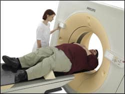
| The Brilliance Big Bore CT from Philips can sustain up to 650 pounds. |
Through his research and experience, Uppot has found that there are practical and technological challenges for each modality, but a good diagnostic image can still be acquired with some adjustments and some new innovations.
Knowing Your Limits
Many radiology centers report turning away obese patients who show up for an exam because they are too heavy for the scanner’s weight limit or too large for the bore hole’s diameter.
Table weight limits for CT and MR scanners are typically around 325 to 350 pounds for most machines, but it can be difficult to determine if an obese patient can fit into the bore just based upon his or her weight.
Conventional bore diameters for MR are about 60 cm and 70 cm for CT. Underestimating or not premeasuring the patient’s girth can potentially halt workflow and upset other patients and referring surgeons.
“In a very busy radiology department, you have patients scheduled every 15 or 30 minutes, and if that train is derailed because the patient can’t fit on the scanner, it becomes a huge problem. The whole schedule now gets backed up,” Uppot said.
Facilities with low- and midfield MRIs with open configurations can be one solution, but the image quality will be less sharp compared with a 1.5T scanner, and there are no such configurations for CTs.
Realizing these challenges, manufacturers have responded with new MRs and CTs with larger bore diameters and increased field of view and tables. For example, Philips Medical Systems, Andover, Mass, has recently introduced its Brilliance Big Bore CT, which can sustain 650 pounds.
Similarly, Siemens Medical Solutions USA Inc, Malvern, Pa, offers its new Magnetom Espree 1.5T MRI with a 70-cm-diameter gantry combined with a 120-cm-long magnet that can help reduce an obese patient’s claustrophobia. In addition, the company recently launched the Magnetom Verio, a 3T MRI system with 70-cm open bore and Total Imaging Matrix technology.
Despite these advancements, technologists and appointment schedulers may still stop an imaging center’s workflow to try to accommodate an oversized patient who meets the table-weight limits but is on the edge of the gantry-diameter restrictions.
For these edge-of-the-bore cases, Uppot recommends that radiology centers invest in a common Hula Hoop, which can then be modified into a “Leo Loop.”
The “Leo Loop,” nicknamed after one of its creators, is a customizable, lightweight, and transportable technologist accessory formed from a Hula Hoop and duct tape. Its purpose is to mimic the MR or CT gantry, thus avoiding technologists from stopping workflow for a wasted attempt to squeeze the patient through. Leo Garcia, an RT at San Antonio Orthopedic Group, and radiologist Richard Benedikt, MD, a partner at South Texas Radiology, San Antonio, developed the concept. (For suggestions on how to create your very own Leo Loop, see the sidebar to this article at right.)
Other Obese Imaging Challenges
Aside from the patient’s size and weight for gantry-based modalities, each technology has its own image-quality issues with obese patients.
X-ray
Attenuation: x-ray beams become attenuated when going through fat. One way to compensate is to increase the kilovoltage peak (kVp) and the milliamperes per second (mAs). The increase will vary by body part. For example, for the typical chest x-ray, Uppot recommends an increase from 90 to 95 kVp to 100 kVp; for the mAs, he recommends the typical 2 to 2.5 mAs to be increased to 4. Uppot notes that these increases will not eliminate the attenuation, but it should improve the image quality.
Motion artifact: The larger the distance that the x-ray beam has to travel, the more chance that there will be motion artifact. Thus, if the x-rays have to travel through extra layers of tissue, there’s a chance that the patient may breathe or move during the passage and result in a blurring of the image. Technologists should take extra care to remind patients to hold their breath and stay as still as possible.
Cassette size: The largest x-ray cassette size is typically 14 x 17 inches, but an obese person’s chest or abdomen can be much larger. If technologists attempt to center the cassette in the area of interest, they may miss or cut off information on the periphery. Uppot recommends that technologists not try to center a single cassette in the abdomen and chest. Instead, he suggests imaging each quadrant separately with four cassettes.
Fluoroscopy
Accurate fluoroscopy is critical for morbidly obese patients because of the growing popularity of laparoscopic gastric bypass surgery. After the surgery, patients perform a postsurgical barium swallow test using fluoroscopy.
Table width and weight limits: The standard fluoroscopic table weight limit is around 350 pounds, but as with CT and MR scanners, manufacturers are now introducing wider tables that support up to 500 to 700 pounds.
Narrow opening between the table and x-ray tube: Standard fluoroscopy systems can have narrow openings between the x-ray tube and the table. Again, manufacturers are now increasing these diameters, some up to 63 cm, according to Uppot.
CT
If the patient can be supported by the table and is able to fit into the gantry, Uppot believes that CT is the best modality for imaging obese patients. An obese person’s fat typically causes the internal structures to be more separated, so radiologists can see all the spaces between them. “You can actually get a fairly good-quality image,” Uppot said.
Switching the mAs to automatic instead of fixed: Similar to x-ray machines, increasing the kVp and mAs can generate a better diagnostic image. Also, rather than having the CT set to a fixed mAs setting, Uppot recommends scanning obese patients with an automatic mAs setting. For very thin portions of the body, the usual fixed mAs may be adequate to obtain a good image quality, but thicker parts of the body can require higher mAs. Consequently, setting the CT scanner’s mAs to automatic will allow the CT to determine the mAs based on the body part being scanned. In addition, slowing down the rotation of the gantry will also increase the effective mAs.
Uppot notes that making these adjustments will slightly increase the radiation dose, but said that radiologists must find a balance between obtaining adequate image quality with the higher settings versus the risk of obtaining a low setting scan that results in poor diagnostic image quality—and, thus, a needless exposure to radiation.
Image cropping: For CT and PET/CT, Uppot cautions technologists in their cropping of the acquired CT images, especially when screening for suspected cancer. He explains, “With obese people, you tend to think that there’s nothing in the borders except the fat, but if you’re worried about cancer and you’re worried about cancer spread, it could spread to the skin and could spread to the subcutaneous fat. So, you have to be very careful about cropping the images and only viewing the cropped images, because there can be pathology on the outside tissues that might not have been seen.”
 |
| Siemens recently launched the Magnetom Verio, a 3T MRI system with 70-cm open bore and Total imaging matrix technology. |
MRI
Coils: While additional phased array body coils may improve image quality, the patient’s size will reduce the space available inside the gantry, and it may be necessary to simply use the standard coils inside the gantry. This will reduce anxiety for claustrophobic patients as well.
Signal-to-noise ratio: Signal-to-noise ratio can be affected by the obesity of the patient. Uppot has discussed this problem with several engineers and physicists but has not discovered a clear answer for exactly why this occurs, but he suspects that there is some kind of attenuation of the radiofrequency signal as it passes through the patient. Uppot said that the latest MRs with increased table-weight limits have somewhat addressed this problem. He also believes that a 3T would be helpful, but is not aware of any 3T system that can support more than the standard 350-pound table limit.
Field of view: When an obese patient exceeds the MR’s field of view, the image can be distorted by wraparound artifact. Consequently, one must adjust the field of view as widely as possible. However, the typical standard field of view on 1.5T systems is from around 40 to 50 cm. MR manufacturers are now beginning to offer scanners with more expanded fields of view, some as much as 60 cm.
Minor burning sensation: Uppot explains that oversized patients squeezed tightly into the gantry can feel a burning sensation in the tissues of the body that are abutting the bore. “It’s not that they have to be dressed clinically,” Uppot said. “It’s just that they feel some minor heating at that site.” To compensate, technologists can use small blankets or pillows as a buffer.
Ultrasound
Ultrasound is perhaps the most challenging modality for obese patients, according to Uppot. That is because the ultrasound waves have to penetrate through the fat, essentially twice—once when the sound wave enters the body, and then a second time when it bounces back and the image is acquired. Uppot points out that not every 250-pound patient will have a poor ultrasound.
Subcutaneous fat versus intraperitoneal fat: Subcutaneous fat will compromise image quality; however, intraperitoneal fat that is inside the abdomen is less of a problem. Consequently, Uppot cautions, “By looking at the person externally, you have no idea where their distribution of fat is. If their distribution of fat is mostly in the subcutaneous tissues, it’s actually going to be very bad for ultrasound, because the ultrasound is going to have to penetrate through some 10 or 12 cm of fat before it gets to the internal structures that you’re trying to see. But a patient who has most of their fat as visceral fat, in those people, the amount of subcutaneous fat in the tissues will be only about a millimeter. So when you put the ultrasound probe on their skin, you have a beautiful image of the liver, because it only has to penetrate through a millimeter of tissue before it gets to the liver.”
Uppot recommends that technologists use the lowest-frequency transducer available, perhaps as low as a 1.5- or 2.0-megahertz probe, which can penetrate more easily through larger fat tissue. In addition, because distance is so important for ultrasound, Uppot suggests technologists image patients on a table and press down on the patient as much as possible, thereby decreasing the distance the ultrasound waves travel.
With the latest trends in obesity showing greater prevalence in America, imaging manufacturers are now responding with new features for oversized patients. However, with DRA 2005 and subsequent reduced reimbursements, it may be some time before this new generation of bariatric imaging equipment reaches its intended patients.
Tor Valenza is a staff writer for Medical Imaging. For more information, contact .


