Historical quote: “If you build it, they will come.”
Historical quote, updated for the modern-day radiology department: “If you build it, they will come” … as long as it increases patient throughput, offers productivity gains, reduces departmental costs, integrates with existing network connections and doesn’t break the capital-equipment budget.
 Kathy Kroupa, chief technologist at Houston’s Fondren Orthopedic Group.
When market research firm Frost & Sullivan (San Antonio, Texas) released its U.S. and European Digital Radiography (DR) Market Report in January, it profiled a market restrained in the short term but positioned for growth in the long term, with predictions for global revenues of $356.1 million in 2007. Since 1998, the first year “significant” numbers of DR systems were sold commercially, global market penetration has been “marginal at best,” the report noted — quite obviously not a shining example of the “build-it-they-will-come” pronunciamento.
As “marginal” as it may be, however, that customer base includes the likes of Avera Sacred Heart Hospital (Yankton, S.D.), Cherry Hospital (Goldsboro, N.C.), Fondren Orthopedic Group LLP (Houston, Texas), Riverside County Regional Medical Center (Moreno Valley, Calif.), The Children’s Hospital of Philadelphia (Pa.), The Mayo Clinic (Jacksonville, Fla.) and The M.D. Anderson Cancer Center (Houston). Despite differences in their size, their resources and their rationales, all seven sites made the move to DR, with each choosing to hook up with a different vendor.
So how has DR made an impact? Let us count the ways.
No. 1: Patient throughput
“A year ago it was rare that we would be done X-raying at 4:30. We close at 6 p.m., but we would be X-raying way after 6 o’clock. We just couldn’t get them in and out fast enough,” Kathy Kroupa, chief technologist at Houston’s Fondren Orthopedic Group, relates one Tuesday afternoon at 4:30. “Our patient volume hasn’t decreased, it has increased, but we have been able to compress the time needed for each exam.”
Increasing patient throughput may not have been at the top of the list of reasons for Fondren to go digital, but it was certainly a welcome development for the orthopedic practice described by Kroupa as an “upscale doctors’ office” built especially for approximately two dozen surgeons and located within the Texas Orthopedic Hospital. “We went digital because we had to,” she says.
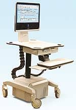
Some of the group’s analog rooms were 20 years old and becoming difficult to maintain and repair, she recalls. Yet Kroupa balked at the chief surgeon’s suggestion that Fondren consider converting to DR: She wanted nothing to do with the only DR she knew — and that was CR (computed radiography). A site visit to the University of Texas Medical Branch (UTMB of Galveston), however, turned her head in the direction of Swissray International Inc.’s (New York), ddRMulti-System, a C-arm configured DR system incorporating CCD (charged-couple device) technology.
Swissray installed its first ddRMulti at Fondren in March 2001, following up with a second installation in June. The practice now images 165 to 175 patients a day in two digital rooms, and it retains one analog room as a backup. Previously, the practice imaged fewer patients in six analog rooms, Kroupa notes.
“We timed a seven-view lumbar spine and a seven-view cervical from start to finish,” she affirms. “With conventional X-ray, it takes about 10 minutes. With digital it takes about five minutes from start to finish.”
The Children’s Hospital of Philadelphia, on the other hand, didn’t doubt that it needed to increase patient throughput: The hospital was ready to extend its enterprise-wide PACS commitment to general radiology; and outpatient imaging, driven by orthopedics, was preparing to expand from a three-room, full-service X-ray area to an additional two rooms that would serve a newly built muscular-skeletal center exclusively. Overall imaging studies were totaling 135,000 with upwards of 70,000 being done via conventional X-ray.
“Our biggest need when we started this process was one of capacity, especially in our outpatient radiology center,” begins Michael D’Olio, a registered technologist and administrative director of radiography. “We had had three rooms that were not able to keep up with capacity, mainly due to our orthopedic service, so subsequently, other services suffered in terms of having access to getting general films. That’s when we began to look at the [DR] technology.”
As the facility began looking at the technology, the Children’s Hospital also paid attention to the development of that technology. That emphasis led the hospital to Hologic Inc. (Bedford, Mass.) and its Epex system, with a flat-panel detector on a reciprocal arm for vertical work and a table for horizontal studies.
In what could be characterized as rapid-fire succession in the world of DR availability, the first of five Epex systems was installed in July 2001. The second followed in August; the third, in November. The fourth was installed in February, with a fifth slated for this month or next.
“We’ve installed those two dedicated muscular-skeletal rooms and have transitioned all the volume we were trying to do in three rooms into two rooms with additional capacity to do more work,” D’Olio remarks, adding that the department in the first five months experienced a 40 percent improvement in patient turnaround time.
“So now we are backfilling, if you will, our outpatient radiology area, outfitting those three rooms with the same kind of equipment. At that point, we will have the capacity to take us fairly far into the future.”
Not unlike the plan at The Children’s Hospital, the move to DR at the Mayo Clinic was the next step in that provider’s effort at creating a full-digital environment — one that involves the Clinic, its own St. Luke’s Hospital and two family-medicine practices, all in Jacksonville, and a third Mayo-owned family practice 30 miles away, in St. Augustine.
“We’re doing 180,000 to 200,000 exams a year at the clinic and of those, about 100,000 can be done on the FD system,” informs Rob Sherrill, director of radiology operations. “What the FD does,” he adds, “is it gives us the ability to keep pace with our growth. We really don’t have the space to put in more radiographic rooms.”
The “FD system” to which Sherrill refers is the Multix FD (flat detector) two-detector system from Siemens Medical Solutions (Iselin, N.J.), also the Clinic’s PACS vendor. Installed approximately 20 months ago, the FD replaced an older, analog room as part of a pilot project to test the FD’s roadability, if you will, prior to full production. The clinic complements the FD with an older Siemens Digiscan 2T chest room and four CR rooms outfitted with FCR 5000 readers from Fujifilm Medical Systems USA Inc. (Stamford, Conn.).
Currently, the Clinic averages 150 studies a day on the Multix FD, 80 of which are routine PA and lateral chests; the remainder are extremities ordered by orthopedic physicians.
Sherrill addresses DR’s impact on patient throughput by explaining the Clinic’s customary approach to imaging — an approach that had been designed with throughput issues in mind when CR burst onto the digital X-ray scene.
Technologists shoot and process CR images, then send them to a QC (quality control) workstation. The techs never see their images; the only time they know something needs to be done is when a QC tech calls and tells them to retake a particular view.
Nowadays, images taken on the Multix FD also flow through to the QC desk, but because the monitor is in the exam room with the system, the technologist knows instantly whether or not the image is a good one.
“It actually speeds up the process because they don’t have to wait for someone to call them and tell them to redo a lumbar view or whatever,” Sherrill says. “So we have seen some efficiencies.”
No. 2: Easy installation, integration
The installation and connectivity experiences of six of the seven sites can be summed up in the words of Jack Slover, radiology administrator at the Riverside County Regional Medical Center (Moreno Valley, Calif.): “Installation went great!”
The seventh, the Mayo Clinic, anticipated some problems since its Multix FD was installed for the purposes of refining workflow and connectivity issues before the model went into full production. As Sherrill recollects: “When we first got it, the system could not run both detectors at the same time. You had to have one hooked up physically, and we had to have the engineers come in and hook up the other detector; the interface wasn’t there. In the beginning it needed quite a lot of refining. But that was the reason they put it here.” Now, the system works without a hitch.
Slover’s exuberant response refers to the installation of a new Philips Digital Diagnost approximately 18 months ago, as part of Riverside’s move into a brand-new building in 1998. The Digital Diagnost, configured as a single-detector in a table and installed in the hospital’s orthopedic clinic, was one piece in a fleet-size purchase of Philips’ diagnostic equipment and PACS network. Open three days a week —Mondays, Wednesdays and Fridays — the clinic performs approximately 13,000 procedures a year, according to Slover. He estimates that the radiology department as a whole is likely to rack up 95,000 procedures in the current fiscal year, ending June 30.
“We had only a small glitch in the beginning. It was sending all images to PACS twice,” he details. “But Sectra Imtec AB (Sweden), who does the software for Philips’ PACS, was out here writing code and it was cured within two or three days.”
The most-oft quoted reason given for a hassle-free installation is communication among the site, the main vendor and any subcontracting parties, with personnel from all entities participating in the project before, during and after installation.
“We didn’t have any surprises,” acknowledges Deborah Powell, radiology manager at Goldsboro’s Cherry Hospital.
But then, Cherry, a 588-bed inpatient psychiatric hospital whose imaging patients include prison inmates and residents of a state-owned training facility for the developmentally disabled, also doesn’t have a PACS or other existing network connections. Its needs were simple and straightforward: Install a DR room and have it print images.
The hospital replaced its seasoned analog system with a Canon Medical Systems’ (Irvine, Calif.) CXDI-22 Universal system purchased through distributor Diagnostic Imaging Inc., a division of PSS World Medical Inc. (Jacksonville, Fla.). However, the package also included workstations from Sunquest Information Systems (Tucson, Ariz.) and a laser imager from Eastman Kodak Co. (Rochester, N.Y.) — all of which needed to talk with the DR system and one another.
“Canon had to get together with Kodak to make sure everything interfaced — and it did,” Powell remembers.
For service and upgrades since the original installation in April 2000, Cherry has relied on local supplier and service provider Blue Ridge X-Ray Co. Inc. (Winston-Salem, N.C.).
“Installation went fairly well,” observes Robin Berke, radiology director at Avera Sacred Heart Hospital, located in the southeast corner of South Dakota.
“The only thing we didn’t expect was that the unit was not intended for printing films and, since we don’t have a PACS, that’s what we’re doing. We’re printing films and you’re rather limited on your [printing] format because of the detector.”
Avera’s unit is a Kodak DirectView DR9000 that was installed last November. The one, fixed C-arm detector is expected to handle 24,000 trauma and general radiography exams a year, the total number of studies done by the two analog systems it replaced.
“It’s working fine for us now,” Berke contends. “We basically have a choice of either filming one-on-one or two-on-one, and that’s our choice. It’s just that we were hoping we would be able to film four-on-one, like when we do an extremity. It’s a waste of film to have to print a single view of a wrist on a 14×17 film.”
A PACS is on the drawing board, he adds, anticipating that his hospital will be part of an enterprise-wide installation connecting all independently run Avera hospitals in Minnesota, South Dakota and Iowa.
Printing also mattered to M.D. Anderson Cancer Center, Houston’s world-renowned cancer treatment facility. And that’s not because Anderson, unlike Cherry and Avera Sacred Heart, has no PACS. It does. In fact, it has two.
“One of the things we had to work with GE Medical Systems (GEMS of Waukesha, Wis.) on was, they had to develop it so we could print to any printer,” offers Reggie F. Munden, M.D., associate professor of radiology and section chief of thoracic imaging.
When Anderson started its DR installation in 1999, it went with a GEMS Revolution XQ/i system, which Munden remembers as GEMS’ first clinical installation, closely followed by a research agreement between the hospital and the company.
“Subsequently, we liked it well enough and were impressed with it well enough that we starting acquiring other ones,” Munden says. “We currently have six units installed.”
The six units are situated throughout the M.D. Anderson complex: one in an inpatient area, one by the emergency center, one in the middle of the diagnostic CT suite, two in outpatient radiology and one in an outpatient imaging center off-site. The six systems conduct an estimated combined total of 350 cases a day, all lateral and PA exams.
Meanwhile, Anderson began its conversion to PACS three years ago — first with a GEMS PACS that the hospital does not use anymore, and more recently with what Munden describes as a “robust” image-distribution system from Stentor Inc. (So. San Francisco). Archives from the two vendors are running concurrently, while Anderson evaluates its PACS options, Munden denotes.
While the DR units feed into Anderson’s need to produce digital images for PACS, the hospital has continued to crank out analog films as well. As a result, Anderson has yet to institute soft-copy reads.
“We were trying to get to centralized printing because we still print images,” elaborates Munden, “which meant we had to get systems that would allow you to transport images electronically to be printed so we needed to get digital imaging onto PACS software for distribution. We also needed to print centrally so we could control QA [quality assurance] centrally.”
No. 21/2: Connections, continued
“We were really surprised that we didn’t [have networking problems],” comments Kroupa, at Fondren. “The gentleman who is in charge of our IS (information services) group, Wesley Spears, is very good at what he does. They sat down with the people from Swissray and mapped out everything before they started on the installation.”
Fondren doesn’t have a PACS network, Kroupa says. Rather, the practice uses an Image Manager System from Swissray that serves the doctors’ offices exclusively. Storage consists of a DVD jukebox with a 12-day memory reserve and retrieval capability: At the end of 12 days, the oldest images are moved onto DVD format.
“The process of networking the [Epex] units to our RIS [radiology information system], to our PACS has gone very, very smoothly,” D’Olio, of The Children’s Hospital, testifies. “The product was already well-developed. [Hologic] worked with IDX Systems Corp. (Burlington Vt.), our RIS vendor, and with Siemens, our PACS vendor, in terms of communication prior to the installation and some conversation afterwards. It has really gone on without a glitch from the first days we installed the equipment. And that’s why we have stuck with the vendor.
“Rarely are you going to pull something out of the box and not have some issues,” he opines. “But some of that is preparing your staff for the changes, working with the vendor on connectivity issues and making sure they’ve been proven at other sites.”
No. 3: Incidental issues
Overall, all sites expressed satisfaction with their choice of DR, indicating that they find the systems reliable, with little or no downtime. Some sites report that film loss and cassette damage are no longer concerns; others assert that supply costs are shrinking — although Riverside’s Slover points out that he now has the new line expense of a service contract; and all appreciate that retakes are nearly nonexistent.
The fact that the systems and their customized worklists enable technologists to position patients, image them quickly and not leave them to process images, is a plus at all sites, but especially at Cherry Hospital where the patient population can be particularly “apprehensive, challenging and pose positioning problems,” Powell says.
Fondren was the only site that had planned to reduce staff as a consequence of installing DR — its tech force went from nine to five — thus saving on personnel costs. Other sites, the Mayo Clinic, for example, reassigned employees, as is in keeping with the Clinic’s culture.
No one registered any complaints — or kudos, for that matter — about DR image quality, although Anderson’s radiologists initially went on record saying they would not accept DR until they felt sure the image quality met the high standards their cancer center required.
Flexibility, due to system configuration, such as the C-arm on Fondren’s ddRMulti, or functionality, such as the ability to change source-image distance settings from 44 to 72 inches on Avera’s Kodak DirectView system, scored points from their users.
No. 4: Talk in the trenches
Several sites indicate they are looking forward to the applications their DR investments will make possible as the technology evolves, PACS proliferate and digital X-ray becomes the rule, rather than the exception.
“I think some of the big pluses are things coming down the pike,” suggests Munden, of Anderson. “The ability to improve image quality as we get better software and better technology, and dual-energy subtraction. We’re going to put that in, and I think we’ll use it all the time once we get used to it. That’s something you can’t do with analog.”
D’Olio, at The Children’s Hospital, seconds that motion.
“I think we are at a great time in radiology; it’s not like just putting in another conventional room and, if you get a lemon, you just sigh and walk away. It isn’t that way anymore.
“You have to be committed to the training, you have to be committed to giving the time and spending the time with the technologists so that they not only accept the new technology, they [also] become expert at it. That’s a change in workflow and commitment. If you’re not willing to do that, I think you’ll find the results are not what you want them to be.
“The payoff at the end is a fabulous one,” D’Olio insists, “but the journey along the way is you have to be committed to change, you have to be committed to evaluating your workflow both pre- and post-installation of the equipment, and you need to be committed to do the measurements before and after to see if you are achieving what your expectations are to achieve. If you are not committed to changing your workflow, you are not going to achieve what you want to achieve.”
Sherrill, at Mayo, weighs in.
“We’re ahead of the game, I believe, because of our ARP [automated radiological practice a/k/a PACS] and the digital patient information,” Sherrill says. “There are a lot of places that do radiology PACS, but they don’t move it outside. We distribute that all the way down. A person can have a CT here in the morning and be sitting down in their examination room with their neurologist this afternoon and see their MR, their CT, their angiogram right on the PC.
“What’s going to happen is, as we grow, as we replace our older systems with the FD, we will not have to add any more radiographic rooms and still be able to put out the same amount of work. We know that’s a fact because we had the same number of rooms in the hospital five years after we started taking it to the digital environment and we’ve doubled our workload. We’re doing twice as many patient exams as we did five years ago and we haven’t added a single room. Without digital, there’s no way we could have done that.”
Riverside’s Slover is more concise, yet there’s no mistaking his appetite for DR.“I don’t have any misgiving about DR at all,” he intones. “My only problem is I don’t have enough of them!”