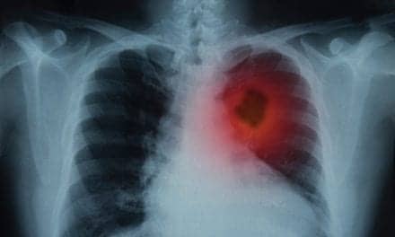
Over the last dozen years, the Santa Monica Cancer Treatment Center (SMCTC), in Santa Monica, Calif, has emerged as a cutting-edge facility for treating cancer patients on the west side of Los Angeles. In doing so, this freestanding center mirrors the state of radiation treatment today, where increasingly sophisticated technology requires better diagnostic imaging support for specific targeting of tumors with stronger therapeutic doses and more specific treatment planning. Through a complement of the latest equipment and successful relationships with nearby clinics, the center is attempting to strike a balance among these factors-a task that sometimes requires the center to be innovative in its methods.
 |
Radiation oncologist Michael Steinberg, MD, and Leopold Avallone, MD, founded the Center in 1988 with the intention of always remaining in the forefront of radiation therapy. Today, SMCTC treats 50 to 60 cancer patients per day and is part of Cancer Care Consultants, a specialized radiation oncology physician group based in Southern California that treats 1,400 cancer patients annually. The center is involved in clinical trials as well as health services research through the RAND corporation and the University of California Los Angeles (UCLA).
Though Avallone has since retired, Steinberg remains on staff in addition to Lisa Chakin, MD, who joined the practice in 1994 and specializes in breast cancer, colon cancer, and prostate cancer. Both Steinberg and Chakin also maintain strong ties with facilities in their area: Chakin as a clinical assistant professor at the nearby UCLA School of Medicine and as a radiation safety officer for the Santa Monica/ UCLA Medical Center and Steinberg as a clinical professor at UCLA and chairman of the cancer committee at Santa Monica/UCLA.
In addition to its skilled staff and community connections, the center is filled with state-of-the-art equipment including high-energy linear accelerator, multiple energy electron beam, computerized tumor localization, a comprehensive computerized patient care management system, computerized three-dimensional treatment planning, a record and verify system, and conformal radiation therapy. The center also offers an ultrasound-based targeting system that is used with external beam radiation therapy to localize targets that may move from one treatment day to the next, and its latest breakthrough? technology is intensity modulated radiation therapy (IMRT), which offers the highest possible radiation dose to tumors while sparing nearby healthy organs, tissues, nerves, and the spinal cord. And according to Jerry Dalrymple, MD, a diagnostic radiologist and medical director of Parkview Imaging Open MRI, also in Santa Monica, IMRT is “the future of radiation therapy.”
“IMRT has the ability to create precision in the shaping of the radiation dose in ways that were previously impossible with conventional external beam radiation therapy,” Steinberg says.
The IMRT system chooses among an array of million of possible radiation beams with varying intensity, rather than using one large beam as in conventional treatment. IMRT thus allows for precision within millimeters. The beam is configured to the specific size, shape, and location of a tumor and can curve to hug concave shapes, an impossibility with 3D conformal radiation. That kind of precision is particularly applicable in brain, head, and neck tumors, and in prostate cancer, though Steinberg says the technology’s application to other tumor sites also is in development.
“IMRT treatments can be used for tumors in the head and neck, brain, prostate, kidney, breast, adrenal, pancreas, esophagus, lung, extremity, spine, and cervix,” he says. “Additionally, any radiation-sensitive tumor occurring anywhere in the body may be a candidate for IMRT treatment.”
IMRT also has emerged as an option to patients unable to tolerate conventional radiation treatment due to side effects or tumor location. The technology’s higher doses of radiation result in potentially higher cure rates. In cases of retreatment, healthy tissue and organs receive much less radiation scatter than that associated with conventional therapy. Finally, the technology offers a reduction in associated costs due to higher cure rate and fewer side effects.
Side Effects Reduced
In terms of the center’s outcomes, IMRT’s use in cases of prostate cancer has resulted in decreased acute side effects such as cystitis, as well as decreased incidence of proctitis and rectal bleeding. Steinberg cites literature from Michael Zelefsky, MD, and his colleagues at Memorial Sloan-Kettering Cancer Center in New York confirming that IMRT in cases of prostate cancer decreases rectal side effects. The study showed that the higher radiation doses (81 Gy) possible with 3D conformal radiation therapy resulted in improved local tumor control (from 55% to 94%) as measured by favorable biopsy compared with lower doses of radiation (64.8-72 Gy). Using IMRT techniques, clinicians were able to deliver these high doses while actually reducing the rate of normal tissue complications from 10% to 2%.1
“However, now we have a technique where we can wrap the radiation around the spinal cord and limit the dose to the cord to prevent the potential development of paralysis,” he says. “We have treated a number of these patients, whom we had to turn away before, so this is a substantial improvement.”
To reap the full benefits of IMRT, the center has had to undergo some changes in the way it does treatment planning, moving from a trial-and-error, forward planning method to an inverse planning approach, according to Steinberg.
“IMRT’s precision gives the treatment planning computers the ability to define complex treatment utilizing inverse planning, where one defines the tumor volume and then asks the computer to optimize the treatment,” Steinberg says. “Prior to this, we always used forward planning, which involved defining the shapes of beams and then trying to modify them. Inverse planning optimizes the beam to the defined target, and modulation of the radiation therapy beam can create radiation dose distributions in almost any conceivable pattern.”
The radiation oncologist thus can designate areas where the radiation dose is not desired, a crucial factor in allowing for higher doses without increased danger to surrounding structures. Because the center now has the delivery methodology to modulate the beam and to move it and the patient during treatment, Steinberg notes that issues also have arisen at the time of treatment concerning whether the tumor or anatomy is static or variable from day to day.
“The way we’re doing treatment planning, it is more intense in terms of physician and physicist input,” Steinberg adds. “This requires many hours of treatment planning.”
The ultrasound-based targeting system was helpful in this aspect for its ability to align the initial complex IMRT position with the daily real-time patient position during treatment. The system creates a stereotactic localization from the original treatment plan that overlays on an ultrasound image, which can precisely define the location of the prostate in real time.
“We are hoping to apply this technology for use in kidney cancer and pancreatic cancer, as well,” Steinberg says. “This gives information to the physician and the radiation therapist to make adjustments on a daily basis.”
In light of these technological advancements, the radiologist’s role in treatment planning with the radiation oncologist also has changed. The shift to more precise methods of treating cancer means a necessary and complementary shift to more demanding imaging requirements.
“The Santa Monica Cancer Treatment Center has more specific imaging needs in terms of their technology and utilizing it to treat patients,” says Jerry Dalrymple, MD, diagnostic radiologist and medical director of Parkview Imaging, which provides imaging services for some of the center’s patients. “As technology becomes more sophisticated, it increases the demands on imaging in terms of quality, spatial resolution, and transmitting the image electronically so that physicians can do their own image manipulation.
“Everybody’s equipment is getting better, and that means the therapist has the ability to focus and deliver radiation with pinpoint accuracy,” he continues. “We have a target, and the goal is to not deliver to any other tissues than that target. The diagnostic radiologist’s role is therefore to provide very accurate information regarding that so-called target. We are using both CT and MRI to provide that information now, and we spend more time going over studies with therapists to make sure the accuracy is maximized.”
The Connectivity Factor
The latter point represents another area where the center has had to exhibit some innovation. In particular, IMRT’s precision is at such a level that the development of a 3D reconstruction of the patient’s anatomy, tumor, and surrounding critical structures became paramount. The center therefore had to create direct links between the places where it obtains images and its treatment planning computers.
“The issue of connectivity between imaging modalities such as CT and MRI is crucial and the biggest hurdle that one has to get over to make these processes work,” Steinberg says. “By gathering these individual images, we can merge them to get the best that CT and MRI scans have to offer, and more precisely define a tumor with critical structures.
“For example in prostate cancer, the use of MRI is particularly accurate in defining the apex of the prostate, and it gives a different view of the remainder of the anatomy as compared to CT,” he says. “Combining information from both gives the most accurate representation of the surrounding structures.”
Currently, SMCTC is linked by T-1 line with the Santa Monica UCLA Medical Center, seven blocks away, from which it receives CT and MR images. In the next phase of the project, the center plans to bring imaging information from a newly installed PET scanner at the Ahmanson Biological Imaging Center at the Santa Monica UCLA Medical Center.
“We’ll be fusing the CT, MR, and PET images,” says Mark Seltzer, MD, an assistant professor of nuclear medicine at UCLA and the clinical attending in nuclear medicine at Ahmanson. “As a functional imaging modality, PET is an excellent tool for evaluating many different cancer types. For instance, in brain tumors, when we are looking for recurrence, the PET scan can show where the tumor occurrence is, whereas anatomic imaging tests can’t differentiate between necrosis and viable tumor. PET therefore can help direct the radiotherapy to those areas. That is the goal for all cancers. Right now, PET has a very high accuracy for evaluating non-small cell lung cancer, colorectal cancer, and head and neck cancer.”
Steinberg says this link would allow for functional imaging and give a whole new dimension to the precise location of the tumor within the patient.
“We can then take that information and infuse it with information from more traditional transaxial images, such as CT and MRI,” Steinberg says. “This has the potential of further precision in defining radiation fields. Already a body of literature is developing having to do with the use of functional imaging in defining radiation therapy fields.”
Although the potential is there, connections between PET and CT and MRI are still in the planning stages.
“Registering the CT images and the MR image with the PET image has been done elsewhere,” Seltzer says. “Other places have lines connected, and we define the specifications that the PET scanner stores and sends. The problem is finding out basically how image data are stored. Is it 8-bit or 16? It isn’t always a simple problem, creating connectivity. Some situations look wonderful on paper and then take months to iron out.”
Facing the Challenges
In spite of its potential benefits, Steinberg notes that adding IMRT and complementary imaging requirements and abilities does present some problems. Initially, there has been no differential in terms of reimbursement for freestanding centers even though the cost difference was substantial.
“There is an increased employee cost due to the number of hours of planning and longer treatment times. It takes longer to set patients up, and it is more complicated and more precise,” Steinberg says.
Hospital ambulatory payment classifications (APCs) do have a reimbursement for the delivery code as well as the treatment planning code, although Steinberg notes that the latter is substantially below the cost of developing the plan. An attempt by the Centers for Medicare and Medicaid Services (CMS) to bundle unrelated radiation therapy and radiology services into the codes also puts a potential roadblock in the development of technology.
“The American Society for Therapeutic Radiology and Oncology (ASTRO) is writing a response letter to the proposed rule expressing opposition to this poorly conceived proposal,” Steinberg says.
“There was no financial benefit to when we added this technology,” he adds. “Those clinics that are only looking at the bottom line aren’t adding this technology, but we believe it is important to have. Not everyone needs it, but we wanted to have it in our armamentarium. The reimbursement issues will clarify themselves. The CMS understands this technology is a revolution in radiation therapy, and hopefully that will be defined by November of this year for freestanding centers.”
Steinberg also notes that ASTRO surveys have found IMRT to be substantially more stressful on the physician and more time consuming.
“The [target] is so refined that we are able to define the field precisely and escalate the dose, increasing the risk of damage to normal tissue if treatment is not planned and delivered in the most eloquent way,” he says. “Treating over a previously treated spinal cord likewise is very stressful for the physician, because if you are not doing what you planned, that can set up the patient for radiation-induced paraplegia.”
The center is undaunted by these issues, however. In fact, Steinberg is looking at increasing the applications of this technology.
“Right now we are looking at almost every tumor system, including breast cancer and head and neck cancer,” he says. “We are going through the planning process for those now. We did about 2 years of research before we stepped in and purchased the equipment initially. Then it took months calibrating the equipment, and treating phantoms to make sure our planning was accurate before we ever put a patient on the machine.
“Even today, each time we go into a new organ system, we go through the same process to make sure there are no trap doors,” Steinberg says. “We are now using the technology in the brain, the head and neck, the prostate, and for cases of retreatment over the spinal cord. We haven’t seen a pancreas yet that was appropriate, but we could do it for those cases as well.”
In terms of imaging, Seltzer points out that technology is advancing to allow many of these different modalities to merge more easily.
“Now we have companies that are making combined CT/PET scanners to be used by radiation oncologists for the same purpose,” Seltzer says. “They are also making it much easier for radiation therapists to utilize the data from all these different modalities, as all of them are complementary. The future is to have all of these together in one machine, and the ability to get instant information.”
“What we will see in 5 years is linear accelerators with CT scan devices associated or built in to reconfirm the location of the tumor and the normal anatomy on a daily basis,” Steinberg agrees.
“Until they are combined, however, we will be using these fusion software packages to provide that anatomic and functional information together,” Seltzer says. “We have not done it yet, but we are in the process of developing that software. It is not that hard to do, but getting connectivity so that the different scanners can communicate is not as trivial as it sounds.”
Elizabeth Finch is a contributing writer for Decisions in Axis Imaging News.
References:
- Zelefsky MJ, Fuks Z, Happersett L, et al. Clinical experience with intensity modulated radiation therapy (IMRT) in prostate cancer. Radiother Oncol. 2000;55:241-9.