|
| Accuray’s CyberKnife System features a linear accelerator mounted on a robotic arm, and is used with the company’s Synchrony Respiratory Tracking System. |
When respiratory gating technology first entered the scene in the 1990s, it gave radiation oncologists a tool to target tumors more accurately. Applying radiation during only the optimal part of the patient’s breathing cycle meant more precise doses as well as more control to avoid irradiating healthy tissue.
But, of course, respiration patterns are not always consistent. No matter how thorough the pretreatment planning, physicians still have to be prepared for unexpected, involuntary patient motion that could affect or even halt treatment delivery. To address this issue, respiratory gating and tracking technologies have evolved to better react in real time to unexpected movement that deviates from the treatment plan.
Monitoring the Patient Position
Pretreatment planning requires determining respiratory patterns ahead of time and tailoring treatment to those cycles. However, variations are almost inevitable when it comes time for the actual treatment. Being able to monitor those variations in real time gives clinicians the data they need on-site to make any necessary adjustments.
In January, Varian Medical Systems, Palo Alto, Calif, received FDA clearance for the patient-position monitoring capabilities of its Real-Time Position Management (RPM) system for respiratory gating and motion management. The system’s cameras track the position of a reflective, plastic marker block placed on the patient’s chest or abdomen. As the cameras track the movement, the system “learns” the motion patterns for that particular area and alerts the clinician to any variations outside the norm.
 |
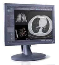
| A brain-treatment plan from Accuray. |
Originally, the RPM system tracked only anterior/posterior motion, but it now analyzes three directions of movement: up and down, forward and backward, and side to side. This change came in response to the realization that most patients can move inside the immobilization devices.
“Some of this motion, like respiration, is periodic, and we’ve been aware of that for a long time,” said Michelle Svatos, PhD, product manager for Varian Medical Systems. “But a lot of motion is not periodic. It’s the patient drifting inside the immobilization device. Maybe it’s internal anatomy that’s shifting around and causing a little bit of ripple on the surface as well, or maybe it’s simply the patient relaxing into a different position.”
Internally, the movement of critical organs can impact treatment. For example, kidney motion can affect how clinicians treat an adjacent pancreatic tumor. “If you were to design a radiation field for only the tumor itself and not take into account the kidney motion that could come directly into the radiation field, you may unnecessarily or inadvertently irradiate the kidneys and cause problems,” said Dwight Heron, MD, chair of radiation oncology at the University of Pittsburgh Medical Center.
Pretreatment planning plays a significant role in successful treatment delivery. The first priority is getting the best possible images of the tumor and surrounding tissue. “Just like surgery, radiation is very local,” Heron said. “If you can’t identify the tumor, you can’t cure the tumor. So, you absolutely need to see it.”
To get the clearest understanding of how the tumor moves, Heron’s team has coupled the RPM system with 4D CT. “3D gives you a shot of the image in time, but it doesn’t give you a sense as to how the tumor is moving in relationship to other structures, say, in the chest,” Heron said. “4D allows us to capture individual images from the CAT scan, and then we can create a movie to actually show what the tumor is doing over the period of the respiratory cycle.”
 |
| Using Varian’s Real-Time Position Management (RPM) sytem, a clinician places an RPM marker block onto a patient’s chest. |
Heron’s team has scanned more than 500 patients using the 4D CT combination, and they have experienced a dramatic drop in margins needed around the tumor: from 15 to 25 mm down to 5 to 7 mm. He has found that performing a second 4D CT halfway through treatment usually reveals identical behavior to the original scans taken 4 weeks previously. “It turns out that it is very reproducible in excess of probably 98% of patients,” he said. This reproducibility means that there will likely be fewer surprises during treatment.
During pretreatment, Heron’s department also incorporates PET/CT into the process, and they plan directly from the PET/CT scan. “When you apply 4D CT with a Varian RPM system to both the CT and the PET, you now have a functional image that’s moving,” he said. “When you fuse them together, it’s a true eye-opener. We’ve discovered things that we did not see with just a 3D PET/CT.”
For example, Heron said that tumors at the periphery of the lung do not necessarily move more than those in the center of the chest, as previously thought. He has also seen lymph nodes in areas that weren’t apparent without the PET/CT. In one case, this method revealed that the center of a pancreatic tumor was more resistant to radiation than the outer areas. “With that information, I could treat the outsides of the tumor with one radiation dose and I could give an extra radiation dose to the center part of the tumor all in the same treatment, using intensely modified radiation therapies,” he said.
Treatments with the RPM system generally last 15 minutes, although this can increase depending on the duty cycle. The RPM technology is particularly suited to large tumors in the abdomen and thorax. It is also popular for certain breast treatments. “Especially for a left-sided breast, you can cause damage to the heart with radiation therapy,” Svatos said. “What they are doing now is gating the treatment and now treating on the inhale position, because when the patient inhales, the lung is blown up, it’s inflated, and there’s more space in-between the breast and the heart, which allows them to miss the heart.”
 |
| The RPM system console depicts the patient and marker block on the left, and the respiratory cycle waveform in the lower right hand corner. |
With more accurate treatment delivery and reduced margins affecting surrounding tissue, patients can take advantage of higher doses in fewer appointments. Lung cancer patients, who often present with comorbidities that further compromise the lungs, particularly benefit. By increasing the dose per appointment, patient appointments could be reduced from 30 to fewer than five. The RPM system allows clinicians to administer these high doses without damaging the surrounding tissue, which is particularly delicate in lung cancer patients. “The normal tissue is especially vulnerable to those high doses,” Svatos said. “Anything you can do to spare the normal tissue and shrink the margin, you’re helping your patient’s odds quite a bit.”
Svatos said that respiratory gating is likely here to stay, because the solution is easy to deliver and is low risk. She adds that one drawback is that the duty cycle can go down and increase treatment time, although not usually as much as one might expect. Still, improvements are in the works—specifically, better efficiency through higher dose rates and improved integration between the optical and x-ray systems.
“One of the things that’s going to go hand in glove with this RPM system is the ability to treat at higher dose rates,” she said. “Another development that’s been coming along with this is the ability to easily and automatically take radiographs—in other words, get internal image guidance when the optical system indicates that something is off.”
Tracking in Real Time
Until recently, respiratory gating seemed to be the best and only option for targeting tumors. Today, an image-guided robotic radiosurgery system from Accuray, Sunnyvale, Calif, automatically and continuously tracks tumors throughout the treatment delivery, without the interruptions involved in respiratory gating.
“It’s definitely superior to traditional respiratory gating,” said Xiaodong Wu, PhD, chief medical physicist in the department of radiation oncology at the University of Miami School of Medicine. “The robot actually moves with the tumor in real time.”
The system combines Accuray’s CyberKnife System, which features a linear accelerator mounted on a robotic arm, with the Synchrony Respiratory Tracking System, which is designed to adapt to patient breathing variations in real time. “What our system does with Synchrony is understand exactly how the patient is breathing and how the tumor is moving,” said Henrik Bacho, director of product management for Accuray. “Then, it moves the linear accelerator in sync with that tumor so that you’re only delivering radiation to the tumor and not to any of the surrounding healthy tissue or critical structures.”
 |
The greater precision comes from the flexibility of the robotic system, which follows the movement of the tumor continuously and in real time. “The robotic arm can give you remarkable freedom of adjusting the position,” Wu said.
To target the tumor, clinicians implant a radiopaque marker, or “gold seed,” inside the tumor itself. “When you take the x-ray image, by looking at the gold seed, you know where the tumor is inside the patient,” Wu said. “At the same time, you use an optic system to monitor the external chest wall motion or the diaphragm motion. So, you have two types of motion, and you build the model to relate these two motions together. During the treatment, you monitor the external motion and project where the tumor will be in real time.”
The system delivers high-dose radiation to very focused areas of the body, and Bacho said the system features sub-millimeter accuracy. “If the patient moves, if they cough, if they roll over, we can track that in real time and adjust the delivery with our robotic system to then match the new patient position and therefore deliver the most accurate dose in radiosurgery treatment available,” he said.
The result is shorter treatment times and better precision. “You can always deliver the right dose to the tumor, and at the same time, you can minimize the dose to normal surrounding structures to protect the normal tissue,” Wu said.
It also means that patients who may not have been eligible for treatment before can now benefit from the CyberKnife with Synchrony combination—for example, if a patient has a tumor adjacent to the spinal cord but has already received the maximum radiation allowed to the spinal cord. “Even though the spinal cord might have received the maximum dose, we can avoid putting any more dose in the spinal cord, and treat a tumor that is growing right next to the spinal cord because of the submillimeter accuracy that the CyberKnife can deliver,” Bacho said.
 |
| Accuray’s Synchrony Vest with LED lights. |
While the monitoring and delivery treatment systems are entirely automatic, clinicians still play a vital role. They prepare pretreatment plans of what to treat and what to avoid, then monitor the treatment-delivery process for any unexpected deviations. “We still need to verify that the model makes sense and that during the whole treatment delivery, everything can be carried out in a safe way,” Wu said.
Taking the Next Steps
Respiratory tracking technology continues to evolve. Last November, Accuray introduced the Xsight Lung Tracking system. The system is designed to target lung tumors without requiring the surgical insertion of fiducials, a common practice that carries some risk of lung collapse and other complications. Removing surgery from the equation speeds up the treatment process, lowers the risk to patients, and ultimately lowers the costs to hospitals.
“[The Xsight] technology enables the system to track a lung tumor directly, simply through the imaging,” Bacho said. “We’ve gotten advanced imaging algorithms that really detect and pull out the lung tumor in the images compared to the surrounding lung tissue, and we are now able to track lung tumors totally noninvasively to avoid the risk of that surgical procedure.”
There are also new implications for the CyberKnife and Synchrony technologies. The current system relies in part on an indirect methodology—monitoring the chest wall to ascertain the position of the tumor itself. Wu believes the next phase will be a direct tracking system, which will involve implanting a transmitter inside the tumor. “From that radio wave, you will know exactly, directly, the motion of the tumor,” Wu said. “And then you will follow the radiation after this transmitter.”
Another possibility involves the use of 4D CT technology, an option that is already in the first development phases. The 4D CT could allow for more precise general tracking.
Wu notes that researchers carefully evaluate the practicality of all these techniques—a criterion that will ultimately decide future trends. “Eventually, the improvement of the precision may not be proportional to the cost itself,” he said. “In this case, we’ll choose what is the most practical and accurate way of doing it.”
No matter the outcome, the technology will continue to address patient motion so that the dose can be delivered more precisely to the targeted area. “Anyone can deliver the right amount to the tumor—that’s not the challenge in radiation oncology,” Bacho said. “The challenge is to minimize the dose outside of the tumor.”
Ann H. Carlson is a contributing writer for Medical Imaging. For more information, contact .