 |
· AAOS Meeting Explores Orthopedic Ultrasound
· Imaging Device Converts 2D Ultrasound Into 3D Tool
AAOS Meeting Explores Orthopedic Ultrasound
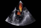
A number of studies presented at the 75th annual meeting of the American Academy of Orthopaedic Surgeons discussed recent findings in the field of ultrasound.

John S. Reach, MD, of New Haven, Conn; James Albert Nunley II, MD, of Durham, NC; and Mark E. Easley, MD, also of Durham, gave a podium presentation on the patient-perceived, surgeon-perceived, and surgically verified diagnostic benefits of enhancing orthopedic physical examination with ultrasonography. Enabling portable, real-time, noninvasive rapid examinations of soft-tissue structures, ultrasound has the capability of soon becoming an orthopedic surgeon’s “stethoscope,” according to the presenters.
In the 1-month study, 64 new patients referred to a Foot and Ankle Service were randomized to an ultrasound and nonultrasound group. Clinical, diagnostic, temporal, subjective, and economic outcomes were compared between the two groups. Results showed that overall time to diagnosis was decreased for patients suffering from peroneal split tears, posterior tibial tendinopathy, FHL tendinitis, and tibialis anterior rupture. One false-positive diagnosis of EHL rupture was made, and there was a decrease in the number of additional studies in the ultrasound group.
Furthermore, 100% of patients indicated they believed that surgeon-utilized ultrasound in the clinic benefited their medical care, and 100% of patients said they would recommend an ultrasound exam to a friend who suffered a similar problem. In 71% of cases, the surgeon believed the ultrasound imaging assisted in final diagnosis.

Another study, presented by Philip Mulieri, MD, of Woburn, Mass; Anthony Festa, MD, of San Diego; Alan S. Curtis, MD, of Chestnut Hill, Mass; and Joel S. Newman, of Ann Arbor, Mich, purported that ultrasound-guided aspiration and corticosteroid injection is a “safe, minimally invasive technique” for treating symptomatic spinoglenoid cysts.
These cysts, which are an atypical cause of shoulder pain, may result in individuals feeling significant distress if they impinge on the supracapular nerve with subsequent atrophy and weakness of the rotator cuff musculature.
Of 18 patients examined, 15 patients noted significant improvement after injection. Seven patients had surgery at an average of 2.6 months after injection, and two patients required cyst decompression. The study authors assert that ultrasoundguided aspiration and corticosteroid injection may be used as an alternative or adjunct to operative management.
—Elaine Sanchez
Tech Zoom
Imaging Device Converts 2D Ultrasound Into 3D Tool
With just a few simple adjustments, clinicians can turn their standard, 2D ultrasound into one capable of producing 3D images and slices.
Approved by the FDA in May, Eigen’s new imaging device, the ei.Nav/Artemis?, allows physicians to select and biopsy a location within the boundary of the prostate with pinpoint accuracy. It is so accurate, the manufacturer actually considers the technology to be 4D.
“The fourth dimension is motion compensation. If the doctor is navigating the prostate and the prostate or the body moves, our software automatically adjusts for that movement. So the doctor knows exactly where he is at all times,” said Michael Castorino, CEO of Eigen. He notes that this level of information is particularly useful for biopsies. “Today, using 2D, it is a blind biopsy. This device will actually direct them to where they want to go, as well as where they plan to pull the tissue, and archive it accordingly.”
This precision means the exact biopsy location can be revisited—or avoided— during repeat procedures. It also generates an image of 3D biopsy coordinates for future reference. Physicians can use the 3D imaging to slice through the prostate, much like what is possible with CT exams.
“Once you have a 3D image, you can slice through the gland and can see all the slices through the prostate,” Castorino said. It is even possible to see a surface-rendered cross-section. “Clinicians will have both the live, 2D image from the ultrasound machine on our monitor, as well as three other panels they can view the 3D imaging on.”
“With as estimated 1.5 million biopsies performed each year, conventional biopsy is blind to cancer, as 20% to 30% of cancers are missed, and detected cancer may not be clinically relevant,” said Al Barqawi, director of research at the University of Colorado Health Sciences Center. “The benefits of advancements in imaging will improve our ability to accurately guide tissue sampling, improve diagnosis, and manage the disease progression. We will be able to treat patients to what they have, not what we think they have.”
All without making any changes to the existing workflow. For clinicians with an existing ultrasound, they lay the existing truss in the cradle that is on the Artemis’ tracker, and they are done, according to Castorino. The system is compatible with ultrasound units from all major manufacturers. Artemis is also available with complete ultrasound functionality for those without an ultrasound unit or for anyone looking to replace existing equipment.
“We designed the product so as to not change any of the existing workflow,” Castorino said. “We’ve had many, many key opinion leaders and doctors as part of our team in developing this product over the last two years, and we were very careful in making sure that it was not adding any more time to their workflow, and that it was actually enhancing efficiency and diagnostic capabilities.”
The first 10 production units are spoken for by the university sites where those contributing clinicians work, including the University of Colorado Health Sciences Center and the University of California, San Francisco. Artemis is available for immediate order in the United States.
In addition to enhancing urologists’ existing ultrasound machines, those who employ the Artemis will be able to dial into a central database of information gathered from system users around the globe.
“There is the ability for us to have every Artemis in the world download anonymized patient data, which means we will be able to track the progress of this machine, and everybody’s patients, with a number of different variables,” Castorino said. Those variables include age, body mass index, and Gleason scores. “We will collect data on a number of different statistics, so that very quickly, the doctors will have a central database that they can go to see how certain treatments in certain techniques are being used by other doctors, and what the outcomes are.”
With regulatory approval under its belt, Eigen is looking toward a myriad of other applications where the Artemis could prove beneficial.
“We will be using this device for therapy and for all of the other difficult anatomies to image, biopsy, and treat, including the kidney, liver, thyroid, and pancreas,” Castorino said.
In addition to helping increase precision in targeted focal therapy and even high intensity focused ultrasound, the system is ideal for conducting targeted brachytherapy, because doctors can actually place the seeds in the exact spot they want them to be in, and go back to that exact spot in the future, should the need arise.
“Since prostate cancer strikes a staggering one in six men, it is startling to me that until now blind biopsies have been the standard of care once cancer of the prostate is suspected,” Castorino said. Other than skin cancer, prostate cancer affects more men in the United States than any other cancer, and nearly 30,000 men in the United States die of prostate cancer each year. “Artemis is intended to give the nation’s nearly 10,000 urologists the technology to navigate to a desired location for prostate biopsy and record this information for future reference, treatment planning, and monitoring.”
—Dana Hinesly