Point Break: A Q&A with Cell>Point?s New President
Product Showcase: Stanford Hospital and Clinics Select Self-Shielding Mobetron for IOERT
University of Wisconsin Installs Inveon Preclinical Imaging System
FDA Clears Nitrogen-Based Cryoablation System from Endocare
Point Break: A Q&A with Cell>Point?s New President
 |
| “99mTc-EC-DG could be a very important agent for those cases where you either can?t afford a PET or you don?t have access to FDG.” ?David Rollo, MD, PhD Cell>Point Inc |
David Rollo, MD, PhD, believes in the potential of molecular imaging to both diagnose and treat cancer. The former chief medical officer of Philips Medical Systems (Andover, Mass), Rollo recently accepted the position of president at Cell>Point Inc (Centennial, Colo), where he will lead the development of molecular imaging agent technetium-99m-ethylenedicysteine-deoxyglucose (99mTc-EC-DG). Medical Imaging spoke with Rollo about the agent, which could image certain cancers and cardiovascular diseases using facilities’ existing SPECT gamma cameras. Cell>Point hopes to submit 99mTc-EC-DG for FDA approval in 2007.
MI: Please describe 99mTc-EC-DG.
Rollo: In the phase I clinical trials and clinical studies that have been done in Asia, 99mTc-EC-DG has a direct affinity for any type of tumor that has increased metabolic activity. It is similar to FDG in that it is an ubiquitous agent. Now, I said “ubiquitous” very specifically, because it doesn’t differentiate between types of cancer. Many molecular imaging agents on the market are target-specific. The problem is, you don’t know—when you have that positive CT or MR—which of the many cancers you have. So, you need something that’s a general cancer-seeking agent, and the only alternative to FDG that I’m aware of is 99mTc-EC-DG. Therefore, it could be a very important agent for those cases where you either can’t afford a PET or you don’t have access to FDG.
MI: What phase has Cell>Point reached in testing 99mTc-EC-DG?
Rollo: The approval for the completion of phase I occurred several months back. Cardinal Health (Dublin, Ohio) has a contract to develop the [good manufacturing process] GMP-manufactured product that will be going into phase II and III clinical trials. We are ready to begin those trials, because we now have a product that can be GMP-manufactured. The trials are anticipated to start sometime in the next several weeks.
MI: Do you anticipate any side effects or reactions in patients from 99mTc-EC-DG?
Rollo: That’s a very good question. Technetium-99m, the imaging agent, has been around for 25 or 30 years, and, of course, there are no side effects from that. The agent itself, 99mTc-EC-DG, is glucosamine. It’s the same agent that people take when they have arthritic pain; you can buy it over the counter. And the size of the dose that would be given is a fraction of what people would take on a daily basis. So, we’re talking about two very benign agents, not to mention the fact that phase I clinical studies clearly showed that there were no adverse reactions. That was a requirement to move to phase II and III.
MI: How do you anticipate the cost of the agent to compare with the cost of FDG? Is it cheaper or more expensive to produce?
Rollo: First, an issue with FDG is that the radioactive agent [fluorine 18] must be produced in a cyclotron, and it has a very short half-life of 110 minutes. Then, you have to do the chemistry of labeling it to the glucose. With that short half-life, you then have to transport the agent to a place that’s no more than 2 hours away. 99mTc-EC-DG comes in a kit form, a little vial that sits on the shelf, and it can sit on the shelf for as many years as you need to keep it. When you’re ready to use it on a patient, you simply inject the technetium directly into the vial, draw out the sample, and inject it into the patient. So, there’s the convenience of being able to do a patient 24/7, as opposed to FDG, where you have to order it the day before. In terms of the average cost to the people who are buying, FDG today is $250 to $300 [per use]. The average cost of the 99mTc-EC-DG kit will be substantially lower than that.
MI: What’s the half-life on the technetium?
Rollo: It’s 61.5 hours. But the 61.5 hours starts when the patient comes in the room, as opposed to the 110 minutes of the FDG, [which begins at the point of manufacture].
MI: So, it’ll probably offer superior illumination.
Rollo: Yes. The other part about technetium is that it comes in a generator. When the patient comes in the room, you go through the process of eluding the generator to take off the technetium right then and there. So, there’s total convenience for the patient.
MI: Does it also have a therapeutic use?
Rollo: The whole area of molecular imaging is extremely exciting because it allows us to do targeted diagnosis. One of the advantages of 99mTc-EC-DG is that it can be labeled with technetium to perform the diagnostic study, and then if the patient does, in fact, have a cancer, you can replace the technetium with rhenium 188. Now, rhenium 188 is a beta emitter, so, in effect, we’d use 99mTc-EC-DG as a guided missile to deliver the therapy directly to the cancer as well as all the metastatic lesions, which means the patient will be receiving target-specific therapy. That’s extremely important, because target-specific therapy doesn’t have the morbidity typically associated with chemotherapy, which targets the whole body. In this case, all you’re doing is targeting the cancer, so it can be done on an outpatient basis, the patient has no side effects, and the efficacy will be very high in terms of treating the primary as well as secondary lesions.
MI: Is this the first time a molecule has been made available for targeted therapy?
Rollo: There are approved agents for target-specific cancer—non-Hodgkin’s lymphoma, for example, has been treated for years with Zevalin, and in that case, it uses indium to look at the biodistribution of the cancer and is followed with yttrium as the therapy agent. It’s the same concept. This is the wave of the future. We will be more accurate in our diagnosis, we will be able to make the diagnosis at an earlier stage, and we’ll be able to provide the cure with noninvasive methods targeting the cancer.
—C. Vasko
Product Showcase: Stanford Hospital and Clinics Select Self-Shielding Mobetron for IOERT
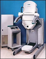 |
| The Mobetron IOERT system from IntraOp Medical is self-shielding and lightweight enough to be fully portable. |
The first fully portable, self-shielding intraoperative electron radiation therapy (IOERT) device has been selected for use by Stanford Hospital and Clinics (Stanford, Calif). The Mobetron, from IntraOp Medical Corp (Sunnyvale, Calif), quickly and safely delivers IOERT to patients; it will be installed in spring 2007.
The Mobetron enables radiation and surgical oncologists to pinpoint the exact area requiring therapy, and it delivers high doses of radiation directly to the affected tissue during cancer surgery. Owing to its portability, the system can be wheeled from one operating room to another, eliminating the need to transfer patients between the surgery and radiation departments.
Conventional linear accelerators can weigh up to 18,000 pounds (9 tons), but the Mobetron’s portability is enabled by its comparatively low weight of just 2,800 pounds (1.4 tons). This weight is achieved by using proprietary 9,000 MHz x-band technology to generate electron beams; most accelerator systems use 3,000 MHz s-band technology, which requires larger and heavier accelerator components. The Mobetron’s maximum electron energy is 12 million electron volts, which offers penetration sufficient to treat 90% of current IOERT patients.
Also, the Mobetron is self-shielding. The device uses a patented energy-control system to eliminate the need for bending magnets, which create significant stray radiation and require substantial shielding to be added to the operating room. The Mobetron design puts the accelerator on an adjustable C-arm system, with a beam-stopper mounted opposite the accelerator to intercept radiation produced in the forward direction.
At Stanford Hospital and Clinics, the Mobetron will replace an Orthovoltage x-ray machine and will be used to treat an array of cancers: colorectal, gynecological, gastric, pancreatic, orthopedic, pediatric, breast, head, and neck. Other US institutions using the device include the University of California at San Francisco, University of North Carolina (Chapel Hill, NC), University Hospital Cleveland, Mayo Clinic Scottsdale, The Ohio State University (Columbus), Clarion Methodist Hospital Indianapolis, and the University of Louisville.
Other options and features of the device are a built-in television-viewing system for treatment-field visualization; extender supports for most surgical tables; an included quality assurance system; bolus for all applicators, including beveled applicators; and sterile caps and drapes.
—C. Vasko
University of Wisconsin Installs Inveon Preclinical Imaging System
The newest preclinical imaging system from Siemens Medical Solutions (Malvern, Pa) has been installed at its first facility, the University of Wisconsin Comprehensive Cancer Center (UWCCC of Madison). The Inveon platform is a dockable, multimodality system, enabling facilities to perform PET, SPECT, or CT imaging, separately or in conjunction with one another.
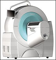 |
| The Inveon preclinical imaging system from Siemens Medical boasts a multimodality upgrade path and a suite of analysis applications. |
“Researchers can expedite the detection and evaluation for preclinical disease treatment by using up to three different scans simultaneously in one compatible system,” said Michael Reitermann, president of the molecular imaging division at Siemens Medical, in a press release. “Inveon will help our customers address research needs, from academic and translational research to drug discovery and development.”
The Inveon system offers researchers the flexibility to visualize and characterize disease, address questions about how diseases progress, evaluate treatments that can slow or stop the transition to cancer in preclinical models of diseases, and transition preclinical studies from the lab to the clinic.
UWCCC—which helped develop the technology used in Inveon through a collaboration between Siemens Medical and Jamey Weichert, PhD, associate professor of radiology—already is using the system’s PET component to take preclinical images. The CT portion was installed in September. The facility will use Inveon for initial preclinical dual-modality virtual colonoscopy studies, as well as for testing of an experimental cancer tracer.
“The Inveon system establishes and delivers the physical limits of PET and CT imaging, pushing researchers to develop molecular imaging agents that are capable of capitalizing on its incredible resolution and sensitivity,” Weichert said. “Within hours of receiving the system, we were able to set up and acquire the first preclinical images.”
—C. Vasko
FDA Clears Nitrogen-Based Cryoablation System from Endocare
A new cryoablation system has the potential to be faster and more user-friendly than conventional argon-based devices. The newly FDA-approved Cryocare CN2 System from Endocare (Irvine, Calif) uses nitrogen, which is more powerful and more widely available in surgical suites than argon, as the freezing and tumor-destroying agent. The form of nitrogen used is called critical nitrogen because it captures the molecule at the crucial point where gas nearly becomes liquid.
Craig Davenport, CEO and chairman of Endocare, explains why argon previously was used in his company’s cryoablation devices. “Previous nitrogen systems were far more difficult to control and, therefore, had a greater potential to cause damage to the surrounding tissue and/or organs that the physician was not attempting to freeze. With high-pressure argon, we also use helium to push the argon gas out of the probes and evacuate it, thereby stopping the freezing effect. So, despite the difficulty of procuring argon and the cost, until recently it was the best gas for cryoablation.”
Paul LaPine, vice president of marketing at Endocare, elaborated on the different freezing agents available. “Both Endocare and Oncura [Plymouth Meeting, Pa] use argon as the principal cryogen,” he says. “However, companies focused on other conditions use other cryogens. For example, most of the dermatology cryo companies use liquid nitrogen. Some of the cardiac companies use nitrous oxide. It really depends on the efficiency level you are striving for.”
There is an important distinction to be drawn between liquid nitrogen, which is considered by many to be inferior to argon, and critical nitrogen. “What makes critical nitrogen so unique is that it will provide more wattage or power with control equal to that of argon,” LaPine says. “The way we accomplish this is a patented method of applying heat and pressure to liquid nitrogen to create near-critical or single-phase nitrogen.”
Adds Davenport, “The cost of nitrogen is about one fifth that of argon, so it will save money for hospitals that own the system and service providers who provide the system. Nitrogen also is available worldwide, whereas argon is not available in many countries.”
Endocare plans to continue marketing its current platform, CS CryoCare, for use by urologists. The new nitrogen system will become its primary platform for use by radiologists, radiology oncologists, and interventional radiologists for ablating liver, lung, and kidney cancers. Currently, cryoablation is being evaluated for the alleviation of bone pain from bone cancers.
—C. Vasko


