Hitting a Moving Target
World?s Largest Proton Therapy Center Opens in Texas
Running the Numbers
Product Showcase: “Drag-and-Drop” Oncology Software to Enhance Workflow
Warm Blanket Decreases Deceptive Brown Fat Uptake in PET Cancer Scans
Electrical Imaging Device Can Detect Tumors Smaller Than 5mm
Hitting a Moving Target
Varian?s respiratory gating technology is used to image, position, and treat
By Dana Hinesly
Although protecting healthy tissue during radiation is a priority, it is often a challenge for tumors in the chest and abdomen, where breathing moves the target by a centimeter or more, depending on the patient. In some cases, accommodating this variance by expanding the margin would require radiation of too much healthy tissue. RPM Respiratory Gating Technology from Varian Medical Systems (Palo Alto, Calif) addresses this issue by synchronizing beam delivery with the patient’s natural breathing cycle.
A noninvasive, computer-aided technique, respiratory gating tracks the patient’s normal breathing pattern and delivers radiation only when the tumor is directly in the path of the treatment beam. Any variation of that pattern automatically switches the beam off, as is explained by Mika Miettinen, senior product manager at Varian Medical, during a recent conversation with Medical Imaging.
MI: How does the technology work?
Miettinen: RPM technology greatly benefits the IG/IMRT [image-guided/intensity-modulated radiation therapy] radiotherapy process when respiratory motion causes the tumor to move. The RPM system is based on an optical marker, called a “marker block,” which is simply placed on the patient’s chest or abdomen. An infrared camera is tracking the motion of the marker and, thus, the motion caused by the respiratory motion can be recorded and provided to the imaging and treatment systems. The correlation between the motion of the internal tumor and the motion of the external optical marker is established using volumetric and/or fluoroscopic imaging in the beginning of and during the treatment course.
 |
The RPM system is used during several phases of the radiotherapy process, including PET/CT imaging, patient positioning, and treatment. All imaging for the treatment planning can be synchronized with respiration, which helps the physician to better analyze the tumor’s motion and provides accurate information for tumor delineation and tumor-margin definition.
Based on the tumor-motion analysis, the clinical team defines gating thresholds, which will be used to gate the imaging and treatment later in the treatment room. After treatment planning is complete and the patient comes in for treatment, the marker block is placed again on the chest or abdomen, with the camera tracking the motion. The patient is coached simply to breathe in a similar fashion as he or she did during the CT-imaging session.
The motion signal is displayed on the RPM workstation and can be viewed and analyzed by a therapist. The gating trigger signal, which is based on the predefined gating thresholds, is automatically sent from the RPM system to the On-Board Imager system, the portal imaging system (PortalVision), and linear accelerator (Clinac).
All imaging for patient positioning, and also treatment itself, is synchronized with respiratory motion, which means the patient is imaged and irradiated only when the tumor location is known with enough certainty. As a result, the patient is positioned more accurately for treatment, and radiation dose to healthy tissue can be reduced significantly in many cases.
MI: How does RPM differ from other methods?
Miettinen: There are other technologies that can be used for this purpose, such as pressure bands placed around the patient’s chest or abdomen, and systems based on spirometers measuring respiration airflow. Varian RPM is an optical tracking system. The optical technology provides some inherent benefits compared to the other monitoring techniques, like the ability to track the motion in space. Also, some systems force patients to hold their breath, but with the RPM system, patients simply have to be able to repeat how they normally breathe. Everything is voluntary in terms of their breathing patterns.
MI: Are there any patients who can’t receive this treatment?
Miettinen: The RPM technology is beneficial in cases where the tumor may move because of the respiratory motion—for example, during lung, breast, liver, or abdominal treatments. Because we place the marker on the patient’s skin, it’s noninvasive and it doesn’t really require any cooperation from the patient’s perspective, so you can use it with every patient. Only one aspect would prevent it from being compatible with a patient. Sometimes a patient’s breathing is so irregular—when they are in a very weak condition, for example—that we cannot trace the motion of the optical marker reliably. In those cases, the treatment tends to be very long because the system waits for the tumor to be in the proper position. In that situation, the gating concept might not benefit the patient.
MI: Assuming patients are able to regulate their breathing, is there any significant increase to the amount of time required for treatments?
Miettinen: It will increase the treatment time, but it only impacts the portion in which we are really treating the patient. Even when it doubles the beam-on time—part of the time the beam is actually on hold—it doesn’t double the whole treatment slot time. If a standard treatment time is 15 minutes, the gating treatment may be 20 minutes. The time increase depends also on the technique used, as RPM supports both the free breathing and the voluntary breath-hold methods.
MI: What technology is required in order to perform treatments?
Miettinen: RPM interfaces with most of GE Healthcare’s CT and PET/CT scanners, and with some other vendors’ CT scanners as well. In terms of treatment, all Varian Medical accelerators are compliant with the RPM technology.
MI: Aside from the ability to spare healthy tissue, are there any other benefits of this type of system for the patient or clinician?
Miettinen: The whole RPM concept is designed to spare the healthy tissue and to treat the tumor accurately to the specified dose. An important design guideline has been patient-friendliness and ease of use. We believe these goals are well met with the RPM system.
MI: What’s next for RPM?
Miettinen: We have a lot of good ideas around it related to the overall motion monitoring, which should be available in the near future.
Dana Hinesly is a contributing writer for Medical Imaging.
World?s Largest Proton Therapy Center Opens in Texas
 |
| The MD Anderson Proton Therapy Center |
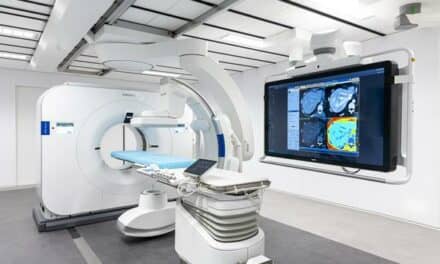
The University of Texas (UT) MD Anderson Cancer Center (Houston) recently opened the doors of its new Proton Therapy Center, a $125 million facility designed to provide the most advanced innovation in radiation therapy. It is the fourth facility of its kind in the country, marking the first time a National Cancer Institute-designated Comprehensive Cancer Center has offered the treatment. More than 40,000 patients at 25 centers around the world have received proton therapy treatment; MD Anderson’s new facility can accommodate 3,500 patients per year, making it the largest in the world.
Proton therapy is a form of precision radiation therapy, minimizing harm to surrounding tissues and optimizing tumor treatment; it has proven invaluable in treating cancers of the prostate, eye, lung, brain, head, and neck. MD Anderson’s Proton Therapy Center will offer external beam radiation therapy treatments for both adults and children. “It’s an important element in the larger practice of radiation oncology at UT,” says Michael Gillin, PhD, chief of clinical physics in the department of radiation physics at UT. “With protons, we are now able to offer another modality.”
In a press release, James D. Cox, MD, head of the division of radiation oncology at MD Anderson, said, “There’s a broad range of patients who will be treated with proton therapy, and they’ll be selected very carefully based on the criteria that their tumors need a high dose and it’s close to sensitive normal organs.”
 |
| Machinery splits the proton from the rest of the atom and sends it to the treatment beam. |
Located on the south campus of UT amid a growing research park, the two-story facility has three gantry treatment rooms, one fixed-beam treatment room, an experimental treatment area, patient and research support areas, a synchrotron, and a beam-transport system.
The center incorporates multiple technologies from an array of vendors. Treatment planning is conducted using images from a GE Healthcare (Waukesha, Wis) CT scanner and the Eclipse treatment planning system from Varian Medical Systems (Palo Alto, Calif). Treatment parameters are uploaded to the EMR system—Mosaiq from IMPAC Medical Systems Inc (Mountain View, Calif). Treatments are delivered using the PROBEAT therapy treatment system from Hitachi Medical Systems America Inc (Twinsburg, Ohio), and patient setups are confirmed with a set of orthogonal radiographs taken with Hitachi Medical equipment. Patient-specific devices are constructed in the proton machine shop using computer-controlled milling machines from Mazak Corp (Florence, Ky).
 |
| A form is made for each patient; this form determines the path of the beam. |
An important role of the Proton Therapy Center will be research and exploration of new ways to make use of the technology, including an investigation into disease sites that could benefit from the treatment. The interaction of chemotherapy with proton therapy also will be explored. “The goal for the center is to offer every patient treated there a clinical protocol,” Gillin says. “One basic protocol is to study the effects of protons on normal tissues. Other protocols are disease specific and will study local control and survival.”
In addition to clinical research, Gillin notes, research to improve treatment planning and treatment delivery will be conducted. One important project will be gating the proton beam to a portion of the respiratory cycle for selected patients whose thoracic tumors move more than 5 mm in the respiratory cycle.
The $125 million facility was built through a collaboration between MD Anderson, Hitachi Medical, investment bank and securities firm Sanders Morris Harris Inc (Houston), and other investors.
Cat Vasko is associate editor of Medical Imaging.
Running the Numbers
36% of patients who received 3D conformal radiation therapy (3D-CRT) to treat their medically inoperable stage I nonsmall cell lung cancer (NSCLC) lived 5 years after diagnosis—compared to 10% who received conventional radiation therapy. In a recent study,1 physicians at MD Anderson Cancer Center (Houston) used 3D-CRT to aim several radiation beams at the tumor to shape or “conform” the radiation to the lung. Between 1978 and 2003, physicians treated 200 patients with radiation therapy; 85 received 3D-CRT, and 115 received conventional therapy. “This study proves that three-dimensional conformal radiation therapy improves outcomes for patients with medically inoperable stage I nonsmall cell lung cancer,” said Ritsuko Komaki, MD, of MD Anderson and a contributor to the study.
Reference
- Fang LC, Komaki R, Allen P, Guerrero T, Mohan R, Cox JD. Comparison of outcomes for patients with medically inoperable stage I non?small-cell lung cancer treated with two-dimensional vs. three-dimensional radiotherapy. Int J Radiat Oncol Biol Phys. 2006;66:108?116. Available at: http://dx.doi.org/10.1016/j.ijrobp.2006.04.015. Accessed September 7, 2006.
Product Showcase: “Drag-and-Drop” Oncology Software to Enhance Workflow
 |
Philips Medical Systems (Andover, Mass) recently announced an upgrade to the Pinnacle3 Radiation Therapy System in the form of its new Model-Based Segmentation (MBS) software for image-guided radiation therapy (IGRT) workflow acceleration.
The MBS software reduces the time it takes to contour tumors and anatomical structures. The software also includes an anatomical library of 3D patient organ-structure models, allowing users to “drag and drop” the models onto patient image data—from which point the software will automatically adapt to each patient’s anatomy. “IGRT workflow is enhanced by allowing clinicians to quickly contour the tumors and organs at risk in three dimensions, then propagate the organs to alternate 4D data sets to help physicians determine the extent of tumor movement within the patient,” explained David Robinson, a certified medical dosimetrist and Philips Medical project manager, in a press release.
The library of anatomical models will grow with the addition of new patient data, enabling clinicians to automatically build a database of information based on regional demographic or clinical practice specialties, a previously unheard-of capability.
The current Pinnacle3 system combines AcQSim3 simulation, Syntegra image fusion, and P3IMRT modules; Philips Medical hopes the addition of MBS software will further accelerate the growth of IGRT.
Warm Blanket Decreases Deceptive Brown Fat Uptake in PET Cancer Scans
New research from the St Louis University School of Medicine suggests that a warm blanket is more effective than beta blockers or valium-propranolol at reducing brown fat uptake during PET scans.
 |
| Medhat Osman, MD, PhD, and other researchers have found that covering patients with a heated blanket before PET scans can reduce the brown fat uptake by up to 30%. |
Accumulations of the imaging agent fluorodeoxyglucose (FDG) in brown fat deposits, which help keep the body warm in cold temperatures, can mimic or mask the appearance of cancer in up to 9% of patients. Animal trials have suggested that pharmacological interventions can be effective 30% of the time at decreasing brown fat uptake, but such methods “aren’t risk free,” explains Medhat Osman, MD, PhD, assistant professor of nuclear medicine and PET director at St Louis University School of Medicine. “Patients with medical problems may face drug interactions. And then you run into logistical problems—who’s going to take the patient home? We believe the use of the warm blanket is twice as effective.”
Osman’s team used an electric warmer to maintain a blanket temperature of 77?. The patients were wrapped in the blanket before contrast injection and during the uptake phase. “I think we may approach elimination of brown fat if we keep patients warm the morning of the exam, and then also when they come to the hospital or the PET center to have the study,” Osman says.
Osman and his team tried the warm-blanket method on patients receiving PET scans during the winter months, when brown fat is often an issue—especially in slender women. Comparing patients who had been warmed before the study with patients who had not, the researchers showed a 62% reduction in brown-fat uptake in the blanket group.
The warm-blanket method creates no risk to the patient and generates no up-front costs. “It’s a simple idea,” Osman admits. “But it worked. And sometimes, that’s all it takes.”
Electrical Imaging Device Can Detect Tumors Smaller Than 5mm
NovaScan LLC (Milwaukee) recently announced that it has accumulated a total of $385,000 to develop proprietary technology to improve cancer detection. The majority of the funding came from private investors, who have contributed $285,000; another $50,000 was obtained through a loan by the Wisconsin Department of Commerce, and the remaining $50,000 came from a grant.
NovaScan’s technology involves functional imaging of breast tissue based on an electrical signature. “Each tissue in the human body has a different electrical property, conductivity, dialectric constant—and if you can measure it, you can distinguish between different tissues, and, more importantly, between healthy and diseased tissue,” says Larry Wells, NovaScan CEO and co-founder. “We’ve measured the electrical properties of different tissues and cataloged fat versus fibrous tissue versus a tumor, as well as the different types of tumors—benign and malignant—and have developed a catalog of those different properties. And that’s the basis for the next step.”
That next step, Wells explains, is to develop a prototype system to scan patients. NovaScan already has entered into an agreement with a Milwaukee-area hospital for a clinical trial on as many as 50 patients. “We’re going to catch them before they get their biopsy,” Wells explains. “Since we’re trying to compare ourselves to x-ray mammography, we’re going to try to match the compression that they had and the views that they had during the mammogram, and then collect our electrical-property data.”
Of course, in the ideal world, the device would work as an adjunct to x-ray mammography, imaging simultaneously and adding as little as possible to total compression time. “The last thing we want to do is materially increase the compression time on the breast. What can the patient tolerate—maybe 3 to 5 seconds? There’s a challenge there. And we have to make sure that our device doesn’t interfere with the acquisition of the x-ray, and vice versa.”
Ultimately, Wells hopes, his company’s electrical imaging will reduce or negate the need for biopsies. “They can be painful, they can be scarring, and they’re expensive,” Wells says. “And they needlessly put the patient through trauma—80% of the biopsies are negative. That tells you where that technology is.”
Electrical imaging can identify tumors smaller than 5 mm, Wells says, and can do so before angiogenesis kicks in to a significant degree—thereby enabling earlier diagnosis. “We want to be sooner, in terms of the genesis of the tumor,” he explains. “If you could, at the screening stage, provide information that now requires maybe a diagnostic mammogram and some other techniques, you could save the patient a lot of time and the health care system millions of dollars.”
Wells stresses, however, that electrical imaging has a long way to go and a lot of difficult hurdles to clear before rendering biopsy a thing of the past. “As far as I know, we’re the only one who’s done this tissue study to the degree that we have,” he notes. “But the literature’s full of people who have electrical-property imaged different tissues. We’ve just taken it a step further. There is a lot of competition out there. But that’s good news for patients.”
—C. Vasko