Trials Begin for PET Agent to Track Tumor Angiogenesis
FDA Approved: Varian Medical?s Trilogy Treatments and Trilogy Tx Cleared by FDA
Running the Numbers
Complications Might Arise Following Balloon Brachytherapy
Study Break
Trials Begin for PET Agent to Track Tumor Angiogenesis
GE Healthcare (Waukesha, Wis) recently announced that it had commenced clinical trials for the first positron emission tomography (PET) agent to track angiogenesis in cancer. The proprietary agent could help physicians identify the location and growth patterns of tumors, providing a more efficient and accurate method of charting the progress of cancer treatment.
Researchers at GE Healthcare first became interested in the molecule’s potential when early animal trials showed that it was more effective at imaging the growth or reduction of tumors than volume reconstruction or glucose metabolism analysis. “The goal is to find out whether we can monitor the tumor—whether it’s progressing or regressing—more quickly than we can using the present methods, which primarily are using CT scans and looking at the size of the tumor or lesion,” explained Don Black, head of research and development at GE Healthcare’s medical diagnostics division.
The agent is a radiolabeled peptide configured to bind to specific integrin receptors, which have limited tissue distribution but high levels of expression during tumor growth. “One of the things that distinguishes cancer cells is that they require a much greater blood supply,” Black said. “So this drug binds to cell surface markers that indicate they’re trying to get more blood supply.”
The clinical trials are taking place at Hammersmith Hospital (London) under the auspices of Imanet, GE Healthcare’s international network of imaging research centers. The first trial measured tolerance of the agent on 10 healthy volunteers. “The radiation dosimetry seems fine,” said David Brooks, MD, chief medical officer at Imanet and head of the trial. “The volunteers haven’t come to any harm that we can detect. Now, we’re starting the patient series to see how well it performs.”
The patient portion of the trial will involve imaging 10 women with recurrent breast cancer before and after cycles of chemotherapy to test the agent’s accuracy at tracking growth and reduction of Stage IV metastatic tumors.
“This is a very novel way of looking at tumors and their growth,” Brooks said. “Eventually, we’ll be interested to see whether it performs better than looking at glucose metabolism, a conventional FDG PET.”
Beyond that, Brooks envisions the agent’s potential to help better evaluate new treatments. “A lot of drugs are now being designed to target new vessel growth by cutting off the blood supply to tumors,” he said. “This is a way not only of detecting tumors, but also of seeing whether these new drugs targeting integrins are actually effective at stopping tumor growth, using imaging as a bymarker.”
—C. Vasko
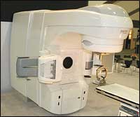
DA Approved: Varian Medical?s Trilogy Treatments and Trilogy Tx Cleared by FDA
 |
| The Trilogy and Trilogy Tx (shown) from Varian Medical are now cleared for use during precision radiotherapy. |
Varian Medical Systems (Palo Alto, Calif) recently announced FDA 510(k) clearance of an expanded range of Trilogy treatments, and of its new Trilogy Tx device.
The Trilogy line of linear accelerators is now cleared for use not only during stereotactic radiosurgery, but also during precision radiotherapy on lesions, tumors, and other conditions throughout the body requiring radiation treatment. Trilogy treatment can take as little as 10 minutes, according to Varian, and is designed to be an outpatient procedure.
“The prior FDA clearance for the Trilogy device was limited to benign and malignant cancers, and arteriovenous malformations,” said Lester Boeh, vice president and general manager of Varian Surgical Sciences. “With this new clearance, physicians can begin to provide Trilogy Treatments for functional legions like trigeminal neuralgia and other noncancerous conditions.”
The company also is cleared to introduce its Trilogy Tx, an image-guided radiosurgery model specifically designed for use by surgeons. A prototype of the new Tx was exhibited in April at the American Association of Neurological Surgeons (AANS) Annual Meeting in San Francisco. Boeh noted that future Tx enhancements will be tailored to surgeons.
Running the Numbers
30% of patients in a recent study had tumors that were missed by mammography and ultrasound but were found to be in the same breast via computer-enhanced breast MRI. In fact, in almost 10% of the patients, tumors were found in the opposite breast via MRI. ?We also found a resulting change in the course of treatment in nearly 25% of patients undergoing surgery for newly diagnosed breast cancer,? said Kathy Schilling, MD, medical director of imaging and intervention at the Center for Breast Care at the Women?s Center at Boca Raton Community Hospital, Florida, and one of the authors of the study.1 Schilling and her research team developed and piloted a software platform using computational clinical imaging techniques for the analysis and display of serial-time MRI.
Reference
- Wiener JI, Schilling KJ, Adami C, Obuchowski NA. Assessment of suspected breast cancer by MRI: a prospective clinical trial using a combined kinetic and morphologic analysis. Am J Roentgenol. 2005;184:878?886. Available at: http://www.ajronline.org/cgi/content/abstract/184/3/878. Accessed July 24, 2006.
Complications Might Arise Following Balloon Brachytherapy
Results of a recent study indicate that patients receiving accelerated-partial breast radiation therapy with a balloon brachytherapy system following a lumpectomy can develop seroma. This fluid retention in the breast can require aspiration and complicate exam monitoring, and, in some cases, it led to an additional biopsy or surgery.
According to Susanne Evans, MD, MPH, lead author of the study and a radiation oncology resident at Tufts-New England Medical Center (Boston), “Seroma is not infrequent after surgery, but it typically resolves itself within a few weeks. However, with these patients, the seroma didn’t resolve.”
The study looked at 38 patients who had a balloon brachytherapy catheter inserted during lumpectomy, some while at Tufts and others while at Rhode Island Hospital (Providence). After the procedure, participants were tracked for an average of 17 months. Of the 38 women, 76.3% developed seroma in the breast; for 68.4%, the seroma persisted longer than 6 months. In 11% of the cases, suspicious asymmetries were detected in the seroma cavity’s lining, and the patients required an additional biopsy or surgery to eliminate the possibility of recurrence.
 |
The study noted that persistent seroma formation was more common in patients with higher body weights, and was less frequent in those who suffered from a postoperative infection.
Alternatives to balloon brachytherapy at the time of a lumpectomy include external beam radiation, which carries risks from increased exposure, or delaying catheter insertion until sometime after surgery.
“We look forward to other studies in which the device is placed postoperatively to determine whether this lowers the rate of seroma formation,” Evans said.
The study, called “Persistent seroma after intraoperative placement of MammoSite for accelerated partial breast irradiation: Incidence, pathologic anatomy, and contributing factors,” was published in the June 2006 issue of the International Journal of Radiation Oncology*Biology*Physics, the official journal of the American Society for Therapeutic Radiology and Oncology (ASTRO of Fairfax, Va). Visit www.redjournal.org/article/PIIS0360301606000848/abstract to read the study in full.
Study Break
By Dana Hinesly
The following brief synopses highlight recently published research.
MR Spectroscopy Significantly Reduces Need for Breast Biopsy
Imaging suspicious breast lesions with MR spectroscopy reduced the need for biopsy by 58%, according to a study published in the June issue of Radiology.
Researchers from Memorial Sloan-Kettering Cancer Center (New York City) treated 56 patients with a total of 57 breast lesions using two modalities. Each woman underwent MRI first, followed by MR spectroscopy. Biopsy was performed after imaging, and results were compared.
At biopsy, there were 31 malignant lesions (54%) and 26 benign lesions (46%). All 31 malignant lesions were diagnosed correctly with MR spectroscopy (100% sensitivity), and 23 of 26 benign lesions were diagnosed correctly (88% specificity).
Biopsy could have been prevented in 23 of 40 lesions of unknown histologic type (58%) if patients had undergone MR spectroscopy during their MRI exam, and all cancers would have been detected.
“All cancers in this study were identified with MR spectroscopy. There were no false-negative results,” said Lia Bartella, MD, lead investigator and assistant professor in the department of breast imaging at Memorial Sloan-Kettering. “With the addition of MR spectroscopy to our breast MRI exam, we found that the number of biopsies recommended on the basis of MRI findings decreased significantly. These results should encourage more women to take this potentially life-saving test.”
According to Bartella, MR spectroscopy is fast and well tolerated, and incorporating it into a breast MRI examination could reduce the number of benign biopsies recommended at MRI—reducing both patient morbidity and unnecessary anxiety, cost, and time for both the patient and the medical staff.
Visit radiology.rsnajnls.org/cgi/content/abstract/239/3/686 to read the study in full.
A Second Look Given to Role of Radiation Therapy in Treating Certain Lung Cancers
Treating certain lung-cancer patients with surgery followed by radiation therapy can improve their chances of long-term survival, according to a study reported in the July 1, 2006, edition of the Journal of Clinical Oncology.
The study employed modern equipment to analyze data from 7,465 patients in the surveillance, epidemiology, and end results database (SEER), a national program that collects information on cancer cases from registries representing 26% of the US population. Of the patients, 47% had received radiation therapy after the surgery and 53% had not.
According to Brian E. Lally, MD, a radiation oncologist at Wake Forest University Baptist Medical Center (Winston-Salem, NC) and the study’s lead author, “Postoperative radiation therapy has failed to demonstrate a survival benefit in the past, likely because previous studies used older equipment.”
He added that additional research is needed to assess the role of radiation therapy and the emerging technology in treatment, in part because of the study’s limitations. Specifically, some information that might impact the results—such as whether the patients had microscopic disease that may have been left behind after surgery—were not available to researchers.
The research found that at 5 years, patients whose disease had spread to the regional lymph nodes between the lungs, and who had received surgery plus radiation therapy, had an overall survival rate of 27%, as compared to 20% in individuals treated with surgery alone.
Researchers were hoping to determine if the advances in radiation-therapy technology would result in improved survival. In all patients, Lally and his colleagues found that radiation treatment did not impact survival. However, in patients where cancer had spread to high-risk regional lymph nodes, survival was improved with radiation therapy.
“For the patients who received surgery and radiation therapy, their survival was better than patients who received surgery alone,” Lally said.
To read the study in full, visit www.jco.org/cgi/content/abstract/24/19/2998.
Two Approaches to Treating Brain Metastases Evaluated
A randomized controlled trial comparing the use of stereotactic radiosurgery (SRS)—both when used in conjunction with and without whole-brain radiation therapy (WBRT)—as treatment for patients with four or fewer brain metastases found that adding WBRT to highly focused radiation therapy does not improve survival for patients with cancer and brain metastases, but it could reduce the likelihood of the recurrence of brain metastases.
The study included 132 patients enrolled at 11 hospitals in Japan between October 1999 and December 2003. Patients were randomly assigned to receive WBRT plus SRS (65 patients) or SRS alone (67 patients).
Hidefumi Aoyama, MD, PhD, of Hokkaido University Graduate School of Medicine (Sapporo, Japan) and colleagues found that the median survival time and the 1-year actuarial survival rate were 7.5 months and 38.5% in the WBRT with SRS group, with a 12-month brain-tumor recurrence rate of 46.8%. For the group receiving only SRS, the median survival time was 8.0 months, the 1-year actuarial was 28.4%, and the 12-month brain-tumor recurrence rate was 76.4%.
Death was attributed to neurologic causes in 22.8% of the patients receiving WBRT plus SRS, and in 19.3% of those treated with SRS alone. There were no significant differences in systemic and neurologic functional preservation and toxic effects of radiation.
According to Jeffrey Raizer, MD, of the Feinberg School of Medicine, Northwestern University (Chicago), who wrote an editorial to accompany the published findings, “Patients who have more than four brain metastases should continue to be treated with WBRT. For patients with four or fewer brain metastases, the combination of stereotactic radiosurgery and WBRT improves local brain control but does not affect survival. Therefore, either mode is a reasonable first choice. ? Whether overall quality of life is positively or negatively affected is unknown, but for patients who might be cured of their cancer, omitting WBRT could avoid long-term neurotoxic effects.”
This study was published in the June 7, 2006, issue of the Journal of the American Medical Association (JAMA). To read it in full, visit jama.ama-assn.org/cgi/content/abstract/295/21/2483.

