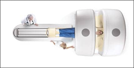
Philips Medical Acquires MRI Leader Intermagnetics
Researchers in Japan Have Performed the First “Helical Shuttle” Scan
Running the Numbers
Oklahoma Hospital Receives First Cantilevered Modular MRI Facility
New Imaging Technique Detects Previously Invisible Smoker Lung Damage
CT Laser Mammography Clinical Results Presented in Milan
Philips Medical Acquires MRI Leader Intermagnetics
In June, Royal Philips Electronics (Amsterdam) announced that it would acquire Intermagnetics General Corp (Latham, NY), which develops, manufactures, and markets high-field superconducting magnets used in MRI. Viewed as a technological innovator in this market, Intermagnetics also provides specialized MRI-compatible patient-monitoring devices and radiofrequency (RF) coils that are predominantly supplied to hospitals.
This acquisition will allow Philips Medical to significantly rationalize its supply chain, enhance its competitive position, and participate in the fast-growing market for RF coils.
In light of the landmark purchase, Medical Imaging spoke to Eoghan O’Lionaird, general manager of MR at Philips Medical Systems, about what impact acquiring Intermagnetics will have on Philips’ market share, as well as how Philips plans on using Intermagnetics’ technology to benefit the medical community.
MI: What percentage of the manufacturing cost of an MRI scanner does the superconducting magnet represent?
O’Lionaird: 40%.
MI: What kind of manufacturing efficiencies do you expect to derive from acquiring Intermagnetics?
O’Lionaird: This is being worked out in a detailed integration plan and will be completed before closing.
MI: What is Intermagnetics’ share of the global magnet market, and how will Philips Medical’s purchase affect Intermagnetics’ other customers?
O’Lionaird: Intermagnetics supplies magnets exclusively to Philips; therefore, the proposed acquisition does not impact other players in the MRI market. Intermagnetics also sells RF coils to Philips, as well as to GE Healthcare and Siemens Medical Solutions. As Philips intends to continue supplying these and other OEMs, the proposed acquisition does not impact other players in the MRI market.
MI: How might this acquisition affect product innovation?
O’Lionaird: We expect innovation cycles to shorten. We also expect that combining Intermagnetics’ leading technology know-how in magnets and coils—as well as other peripherals, such as CAD and fMRI—with Philips’ system design and clinical applications will deliver significant innovation in the future.
MI: How important to Philips Medical are Intermagnetics’ MR-compatible patient-monitoring devices?
O’Lionaird: Philips currently brings Intermagnetics’ MR-compatible patient-monitoring products to market, and we expect that they will be an integral offering to our customers in the future. Intermagnetics brings differentiated know-how and technology in MR-compatible acute care monitoring. As MR becomes more and more used in interventional procedures—using MR real-time guidance for catheterization, RF ablation, ultrasound ablation, electrophysiology, as well as other cardiac interventions—these products, which can be used in the MR suite, will become even more important and valuable to our radiology customers.
MI: With the recent broadening of indications for pacemakers by CMS, do you expect this part of the company to grow?
O’Lionaird: Yes.
MI: How strong is the RF coil piece of Intermagnetics, and what are Philips Medical’s plans for this segment of the business?
O’Lionaird: Intermagnetics is recognized as a leading innovator in RF coils. We expect to leverage this leadership, in combination with the clinical application leadership of Philips, to bring innovative solutions to our customers in the future. Clinical applications are only as useful as the data sets—collected by means of the RF coils—on which the applications perform some functions. The design of the coils as an integral part of the MR system is key, and we anticipate that the integration of the Intermagnetics RF coil capabilities into Philips Medical will create a truly powerful combination.
Furthermore, as MR functionality is extended beyond [hydrogen] proton imaging to include imaging of other nuclei, such as carbon and phosphor, new and sophisticated coil designs that preserve signal and deliver high-resolution data quickly will become increasingly important.
Finally, as MR functionality continues to mature, and resolution and speed continue to be expected—already, MR can image to a single-molecule resolution, even at mainstream 1.5T field strength—higher channel-count coils with ever-more-sophisticated designs will be needed. (Eight channel to 16 to 32 are current; 64 through 128 to quasi infinite channels are the trend.) The capabilities that Intermagnetics brings also will, in this context, be critically important and valuable.
Researchers in Japan Have Performed the First “Helical Shuttle” Scan
Researchers at Kinki University Hospital (Osaka, Japan) have performed the world’s first “helical shuttle” CT scan, according to an announcement from GE Healthcare (Waukesha, Wis). In one scan, Takamichi Murakami, professor and chairman of the department of diagnostic and interventional radiology at Kinki, was able to perform both a dynamic CT angiography and a full organ-perfusion study—with no compromise to temporal resolution.
The new technology provides real-time controls for “shuttling” the CT table back and forth during scanning; it also allows wider coverage for complete organ imaging, up to 120 mm longitudinally. For example, in the images shown here, the liver was covered 22 times (11 round trips), and the maximum speed was 110mm/second. Anatomically adaptive acquisition could potentially be a powerful diagnostic tool for noninvasive clinical studies, the company claims.
|
|
|
|
||||
|
These volume-rendered helical shuttle dynamic images show the entire volume of a liver(160 mm) as the contrast flows into major vessels and perfuses into the liver tissue. Dynamic contrast flow has the potential to provide additional information for the assessment of liver tissue with implications to the management of the patient. The images (from left to right) were captured 1.5, 4, 9, and 16 seconds after the contrast injection. Images courtesy of GE Healthcare. |
|||||||
“The underlying technical concept behind helical shuttle was to break through the old paradigm that helical scans must be performed at a constant table speed,” said Brian Duchinsky, general manager of global CT at GE Healthcare. “This breakthrough is enabled by an innovative, real-time scan-control architecture. Additionally, our engineers have been creating new reconstruction algorithms that can produce images acquired with helical shuttle without artifacts previously associated with varying table speed.”
Murakami added, “Helical shuttle could open up new clinical opportunities, such as ?dynamic blood flow,’ which can potentially offer additional clinical information, leading to a new concept called 4D CT angiography. This additional information could allow us to investigate in detail the relation between tumors and feeding arteries. Furthermore, this information could prove to be very valuable in helping clinicians determine the adequate treatment choice: ablation or medical therapy.”
Murakami also noted that today, “CT perfusion studies are limited to small organs or organ segments only. Helical shuttle holds promise to perform whole-organ angiographic and physiological assessment in a single scan. The possibilities brought forth by this clinical capability could enhance the way we use CT beyond anatomical assessment.”
Running the Numbers
54% of respondents to the Medical Imaging monthly Web Poll say they have a 64-detector CT scanner in their facility. We asked readers who work in facilities that offer CT to name the highest number of detectors on one of their scanners. Here are the answers:.

|
Oklahoma Hospital Receives First Cantilevered Modular MRI Facility

|
| HCG Building Technologies installed a cantilevered-designed modular MRI suite at Cushing Hospital. |
HCG Building Technologies (Ceres, Calif), a provider of hospital-grade modular facilities, recently announced the opening of its first cantilevered modular MRI facility—a custom-designed second-story addition to Cushing Regional Hospital (Cushing, Okla). Cushing Hospital, a division of the Hillcrest Healthcare System, now hosts its first in-house MRI service above its emergency department (ED).
To deal with the hospital’s space constraints, HCG employed a cantilevered design for a portion of the modular building. Also known as a projecting structural design, a cantilever allows constructions above a first floor with support at only one end. In Cushing Hospital’s case, the MRI facility was installed onto the southwest side of the hospital with a 5-foot extension.

|
| The finished product allows the MRI suite to be close to both the ED and the radiology departments. |
“The HCG design team worked hard to accommodate the complex siting requirements and specialized space needs at Cushing Hospital,” said Brad Fallentine, vice president of sales and marketing at HCG. “The cantilever design used empty space above the ED while creating an efficient and comfortable diagnostic area for their patients, staff, and physicians.”
Designed to provide direct access to the adjacent radiology department, the cantilevered suite was commissioned to house the hospital’s first permanent MRI system, an Intera 1.5T from Philips Medical Systems (Andover, Mass). Prior to purchasing the Intera, Cushing Hospital had been relying on a mobile MRI system.
“Once it became evident that we needed an MRI system on a full-time basis, it was important to keep our MRI service within the radiology department, for both convenience and safety,” said Steve Rodewald, director of radiology at Cushing Hospital. “The modular option helped meet these needs, along with our short time line and desire for minimal on-site disruption. And the unique design met our space-constraint issues.”
New Imaging Technique Detects Previously Invisible Smoker Lung Damage
Researchers at the University of Wisconsin?Madison have developed a new imaging technique that exposes lung damage in smokers—even healthy smokers with no other indications of tobacco use.

|
| This image shows a helium image of the lung airways (left) juxtaposed with a conventional MRI (right), illustrating the marked difference between the helium MR image that evaluates the air space of the lungs compared to conventional MRI, which shows images that are proportional to the body?s water content. Note that because the lungs are largely air space, they are not well seen on conventional MRI. |
Although MRI is traditionally used for high-resolution visualizations of soft tissue because of its ability to register differences in water content, it also can be employed in detecting helium, a harmless gas. The research team at UW?Madison, led by Sean Fain, PhD, assistant professor of medical physics, had their subjects—eight nonsmokers and 11 healthy smokers—inhale helium before MR imaging.
The team’s theory was simple: Smoking-related lung deterioration, particularly emphysema, is often characterized by the breakdown of alveoli. With fewer functioning alveoli to contend with, a gas like helium would be freer to move through the lungs. MR imaging upheld the theory, showing that the “diffusion coefficient,” or movement of helium gas molecules throughout the lungs, was related to how much the patient smoked, with increased movement indicating a higher level of damage. CT, which is more frequently used to image the lungs, showed no such correlation.
“It’s one thing to see a disease that already was diagnosed, but another to see changes that no one predicted were there,” Fain said. “This approach allows us to look at lung microstructures that are on the scale of less than a millimeter. Our technique is potentially more sensitive than established techniques. This is the first time structural changes have been shown in the lungs of asymptomatic smokers.”
The researchers believe helium-based MRI scans could have a variety of lung-imaging applications, from measuring the efficacy of experimental drug therapies designed to reduce smoker lung damage to screening for genetic predispositions to emphysema and other lung diseases.

|
| Images of the lung air spaces were derived from subjects in Fain?s study. These images depict typical defects (arrows) in a smoker with 14 pack years on the upper left, a healthy nonsmoker on the upper right, and a smoker with 39 pack years on the lower left. These defects represent regions where the gas is obstructed by narrowing of the lung airways. |
CT Laser Mammography Clinical Results Presented in Milan

|
| On this 56-year-old patient with no clinical findings, the screening mammography in the right breast reveals Tabar type 3 pattern. Near the nipple in the upper outer quadrant, a small nodule is seen (also in compressed view). Using CTLM, angiogenesis is seen centrally and extending into the lower outer quadrant of the right breast. Pathology reveals Invasive Ductal Carcinoma, Grade II?III. |
Clinical results obtained using a CT laser mammography (CTLM) system developed by Imaging Diagnostic Systems Inc (IDSI of Fort Lauderdale, Fla) were featured at the Eighth Annual Milan Breast Cancer Conference, held June 21?23 in Milan. This year’s meeting, “Innovation in Care and Research,” was dedicated to developments in breast-cancer prevention, detection, and treatments. According to IDSI, the company’s clinical collaboration sites provide imaging results from some of their cases using CTLM (shown here).
IDSI’s CTLM system is the first patented breast-imaging system to use laser technology and algorithms to create 3D images of the breast, thereby allowing noninvasive breast imaging without radiation exposure or breast compression. CTLM images blood flow to the breast and could, therefore, be used to visualize tumor angiogenesis. It also penetrates easily through breast implants or dense tissue, unlike mammography, and has been shown to increase sensitivity and specificity of breast-cancer detection.
The company boasts four CTLM systems installed throughout Italy; the European Institute of Oncology, which organizes the Milan conference, is one of the company’s collaboration sites. IDSI awaits FDA 510(k) premarket approval for the CTLM to be used as an adjunct to mammography. However, the system has been approved for marketing in Canada and China, and it has received FDA export certification.

|
| This 62-year-old patient with no clinical findings has a mammography of the left breast that reveals a 19- x 10-mm lesion at 9 o?clock posteriorly, classified as BIRADS 5. The CTLM study, however, demonstrates extensive angiogenesis at 9 o?clock posteriorly. Pathology reveals Infiltrating Ductal Carcinoma grade II, positive estrogen and progesterone receptors. |

|
| This 62-year-old patient presented with a skin indentation at the border of the outer upper quadrant. A mammography of the left breast, Tabar type 3, shows a 15- x 15-mm nodule in the upper outer quadrant; however, the CTLM in the left breast area shows angiogenesis at the 3 o?clock position. Pathology reveals Invasive Lobular Carcinoma. Shown here is the cranio-caudal view. |


