ARRS Report Roundup
Fully Integrated Lung CAD Solution Launched
Running the Numbers
Companies Collaborate with Hospitals for High-Field Body and 128-Channel MRI
FDA Approved: The FDA Clears Two CT Scanners that Provide Increased Comfort and Precision
First Biograph 64-Slice PET/CT System Installed at Emory Crawford Long Hospital
ARRS Report Roundup
by Dana Hinesly
Technological advances fueled interesting research findings presented during the Annual Meeting of the American Roentgen Ray Society (ARRS of Leesburg, Va), held April 30?May 5 in Vancouver, BC. What follows is a primer on some of the key findings released at the meeting.
MRI Studies
- High-Powered MRI Compares with Arthroscopy for Shoulder Injuries. Researchers from the Neuroskeletal Imaging Institute (Merritt Island, Fla) reviewed the shoulder MRIs obtained with a 3T magnet of 100 consecutive patients and discovered MRI had a high correlation with what is found at surgery. Researchers believe the study demonstrates that diagnosing tears can be done satisfactorily without arthroscopy, sparing many patients from surgery. Lead author: Thomas Magee, MD
- MRI-Guided Technique Speeds Fibroid Treatment. Fibroids can be safely treated in 60% less time using manual interleaved MR-guided focused ultrasound (MRgFUS), according to researchers from the Lahey Clinic (Burlington, Mass). Follow-up visits found the new MRI-guided technique produced no serious adverse side effects, and 12 of the 14 patients had marked improvements in their symptoms. Lead author: George A. Holland, MD
- Noninvasive Staging of Fibrosis Possible with Combined Contrast-Enhanced MRI. A study conducted at the University of California, San Diego, found that accurate noninvasive staging of liver fibrosis in nonalcoholic fatty liver disease (NAFLD) was possible with combined contrast-enhanced (CCE) MRI. The team used two contrast agents with complementary mechanisms—iron particles to darken the background liver and gadolinium to brighten the fibrosis—and deemed the procedure repeatable and possibly more accurate than any other noninvasive method for assessing liver fibrosis. Lead author: Claude Sirlin, MD
- MR Cystography Potential Screening Exam for Ureteral Reflux. Researchers from Changi General Hospital in Singapore and the University of California, San Francisco, teamed forces to take a closer look at patients with VUR detected by conventional X-ray cystography who also underwent MR cystography. According to the study, the two methods were concordant for 80% of the ureters and the remaining 20% was split, with MR cystography detecting VUR in one ureter and X-ray cystography detecting it in the other. Lead author: Hui-Seong Teh, MD
CT Studies
- 64-Slice CT Angiography Proves Highly Accurate for Detecting High-Grade Artery Stenosis. Multidetector CT angiography (MD-CTA) holds its own against color-coded Doppler sonography, power Doppler, and B-flow ultrasound in detecting and grading extracranial internal carotid artery (ICA) stenoses in which the artery is closed between 70% and 90%. When performing both MD-CTA and vascular ultrasound exams on 37 patients with 43 known or suspected extracranial ICA stenoses, the team at the University of Munich-Grosshadern (Munich, Germany) achieved “excellent” visualization of vessels in all cases. Lead author: Dirk Andre Clevert, MD
- Body CT Readings with Coronal Reformatted Images Are Faster. Radiologists could interpret 30% to 40% less images per case if they replace axial images with coronal multiplanar reformatted images for interpretation in the multidetector CT (MDCT) evaluation of the GI tract, according to a new study from Emory University School of Medicine (Atlanta). When analyzing MDCT scans for 50 patients referred for GI tract imaging, researchers discovered that the average duration was shorter and confidence was higher when interpreting coronal reformats. Lead author: Sunit Sebastian, MD
- Common Interventional Radiology Procedures Can be Performed at Lower Dose. Safe and effective interventional radiology procedures are possible even when the radiation dose is reduced by as much as 88 percent, according to researchers at Boston University Medical Center. Analyzing CT-guided biopsy, needle aspiration, or catheter placement procedures conducted on 291 patients, the researchers found it possible to accurately perform such image-guided interventional procedures even with slightly “muddy” images. To perform the procedure successfully, clinicians began with a very low dose, increasing it until the procedure is feasible. Lead author: Brian Lucey, MD
- Image-Guided Biopsy Can Prevent Unnecessary Kidney Removal. After reviewing 153 kidney biopsies in 126 patients, researchers at the University of Michigan (Ann Arbor) discovered that the course of treatment was changed for more than 60% of patients. As many as 75 unnecessary kidney removals potentially were avoided. Employing percutaneous image-guided (CT or ultrasound) biopsy of renal masses increased the odds that clinicians were sampling the correct tissue, while simultaneously decreasing the risk of damage to adjacent structures. Lead author: Katherine Maturen, MD
- Kidney Removal for Suspicious Masses Might Be Limited with Image-Guided Biopsy. A closer look at image-guided kidney biopsy was performed at Massachusetts General Hospital (MGH of Boston), establishing the procedure’s ability to diagnose benign kidney tumors with a low rate of complications. Researchers performed CT and/or ultrasound to conduct 407 image-guided kidney lesion biopsies in 377 patients. The study found it was safe to biopsy tumors smaller than 4 cm, according to the researchers, who hope renal biopsy can help prevent unnecessary surgery. Lead author: Anthony Samir, MD
- Combining Different CT Views Gives More GI Tract Information. Improved diagnosis of gastrointestinal (GI) tract problems can be realized by combining transverse images and coronal multiplanar reformats from CT scans, according to research conducted at the Emory University School of Medicine (Atlanta). Analyzing routine abdomen-pelvis CT scans from 50 patients with a simultaneous review of coronal and transverse images revealed 281 lesions; examining transverse images only detected just 259 lesions. Lead author: Sunit Sebastian, MD
- New Device During Coronary Angiography Minimizes Radiation Exposure. A physician at the University of Maryland Medical Center (Baltimore) employed a newly developed extension bar during coronary angiography that allows physicians to remain behind a lead plastic shield yet still be able to control the exam table movement. Using this method, the radiation exposure to the physician’s head, arms, and legs was reduced 90%. Technique developer: Martin Magram, MD
- MDCT Shows Promise for Detecting Bladder Tumors. Bladder tumors can be readily detected using MDCT urography—both with and without contrast—researchers at the University of Michigan Health System (Ann Arbor) found. In looking at 92 patients with bladder cancers, the study found that MDCT was able to identify 87 of the 92 tumors. The study also discovered that it didn’t always matter whether contrast was used or not. Lead author: Jonathan Willatt, MD
- Lower Doses for Abdominal CT of Children Don’t Impact Image Quality. Researchers at the Children’s Hospital of Philadelphia found that abdominal CT radiation dose can be reduced by 60% without compromising image quality. When reviewing abdominal scans from 74 children researchers found that, on average, modulating the tube current to account for body symmetry reduced the radiation dose by 15% over the usual weight-adjusted dose. Lead author: Soroosh Mahboubi, MD
- Latest CT Technology Improves Diagnosis in Emergency Department (ED). Radiologists at Brenner Children’s Hospital (Winston-Salem, NC) are using 64-slice CT and both 2D and 3D images to assess and treat trauma patients in the hospital’s ED. Employing 2D or 3D imaging makes it easier to locate any ruptures and define the full extent of injuries, providing surgeons with the information they need when deciding whether or not to operate. Presenter: Craig Barnes, MD
PET/CT
- PET/CT Better at Detecting Ovarian Cancer than PET or CT Alone. Researchers at Massachusetts General Hospital (Boston) reviewed 54 body CT, PET, and PET/CT examinations on 53 patients with ovarian cancer who were being evaluated for tumor recurrence and discovered that PET/CT’s accuracy (49/53, 92%) was higher than either CT (44/53, 83%) or PET alone (41/53, 77%). Lead author: Sunit Sebastian, MD
- FDG-PET Accurately Assesses Tumor Destruction After Radiofrequency Ablation (RFA). Researchers at Newton-Wellesley Hospital (Newton, Mass) reported that FDG-PET provides more valuable information about the amount of tumor destruction after RFA than CT alone. Clinicians using PET can see which tumor cells are still alive, making it easier to assess how much residual active tumor remains. Lead author: Jennifer Daly, MD.
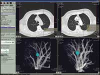
Fully Integrated Lung CAD Solution Launched
Vital Images Inc (Minneapolis) and R2 Technology Inc (Sunnyvale, Calif)—the latter of which recently was acquired by Hologic Inc (Bedford, Mass)—have partnered to launch the Vitrea solution featuring ImageChecker CT Lung CAD software. Designed with the thoracic clinical workflow in mind, R2’s lung CAD solution (ImageChecker) improves the detection of solid lung nodules during review of multislice CT exams, and the integrated solution improves workflow efficiency in lung analysis on Vital Images’ Vitrea workstation.
 |
| R2 Technology’s FDA-approved (PMA) ImageChecker CT Lung CAD System helps detect actionable lung nodules during review of multislice CT images to help minimize observational oversights. This tool is fully integrated into the Vital Images Vitrea 3D workstation lung-analysis application to help streamline workflow. |
“The increased number of thinner and thinner lung CT images generated from thoracic multidetector CT examinations has increased the demands on the clinical radiologist and has mandated significant workflow efficiencies,” said Ella A. Kazerooni, MD, professor and director of cardiothoracic radiology at the University of Michigan Health Systems (Ann Arbor). “We now can easily and consistently integrate CAD findings into the exam workflow via Vitrea.”
Vitrea creates 2D, 3D, and 4D images of human anatomy from CT and MRI data. The integrated CT lung CAD solution includes automatic detection of pulmonary nodules through AutoPoint, which provides automatic temporal comparison of pulmonary nodules, pulmonary artery patency examination (PE) detection software, and a flexible but consolidated reporting feature scalable to a clinical enterprise.
R2’s ImageChecker CT Lung CAD is an FDA-approved system for the use of CAD in improving the detection of actionable lung nodules during review of multi-slice CT chest exams. Its CAD algorithms examine a CT chest study and automatically mark areas warranting further examination, thereby enabling increased diagnostic accuracy and physician productivity.
“Now, Vitrea customers can benefit from the full power of R2’s CT Lung CAD solution in a user interface with which they are familiar from other Vitrea applications,” said John Pavlidis, R2’s president and CEO. “With already more than 100 CT Lung CAD installations, this new integrated solution will only help to increase our CAD’s adoption rate among health care providers.”
The technology has been FDA cleared and was made available April 2006.
Running the Numbers
1,460 64-slice CT scanners have been fully installed in the United States and are in clinical use.* The numbers include 550 installations of the LightSpeed VCT line from GE Healthcare (Waukesha, Wis), 400 installations of the Brilliance line from Philips Medical Systems (Andover, Mass), 320 installations of the Somatom Sensation line from Siemens Medical Solutions (Malvern, Pa), and 190 installations of the Aquilion line from Toshiba America Medical Systems (Tustin, Calif).
* Numbers are based on a June 9 vendor poll.
Companies Collaborate with Hospitals for High-Field Body and 128-Channel MRI
Two companies have announced collaborations with hospitals for the development of MRI technology. GE Healthcare (Waukesha, Wis) and the Mayo Clinic (Rochester, Minn) have announced a program for the clinical development of high-field MRI of the body, and Massachusetts General Hospital (MGH of Boston) and Siemens Medical Solutions (Malvern, Pa) have developed a prototype 128-channel MRI system based on Tim (Total imaging matrix) technology.
The GE Healthcare collaboration is designed to help realize the full potential of 3T MR systems as a diagnostic tool, particularly for the abdomen, heart, breast, and musculoskeletal system. Along with the Mayo Clinic’s Body MRI Advanced Development Unit, GE Healthcare will develop and apply clinically viable techniques and protocols for 3T, allowing Mayo’s patients to benefit from the potential to accurately diagnose such conditions as breast and prostate cancer, liver disease, and coronary artery disease.
“Accurate and early diagnosis is the critical forerunner to effective medical treatment, which is why Mayo is focusing on improving the diagnostic capabilities of advanced MR imaging,” said Kimberly
 |
| The Magnetom Trio 128-channel prototype from Siemens Medical at MGH opens doors to new applications, including abdominal, cardiac, spine, whole-body, and orthopedic MRI at 3T. |
K. Amrami, MD, head of the body MR imaging practice at Mayo. “Both Mayo and GE Healthcare bring exceptional resources and enthusiasm to the development of clinical applications for this exciting new technology.”
The Siemens Medical collaboration involves research and development of the Magnetom Trio with Tim 3T MRI system. The prototype of the system is based on the 102 x 32 Tim architecture—102 coil elements integrated in 32 independent radiofrequency (RF) channels—that has been expanded to 128 independent RF channels and coil elements. The principles and potential applications of 128-channel MRI scanning are being evaluated at MGH, with initial findings showing the potential to exceed current standards of image resolution and parallel imaging, with up to a 25-fold increase in speed.
The prototype is designed to be suitable for clinical and research imaging, especially in cardiology and advanced neurology, where speed and sensitivity are imperative. Cardiac imaging could be conducted in a single breath hold and viewed in real time without radiation or drugs, making MRI a key resource for medical professionals and their patients. With the increase of channels, it is possible to see a shift to rapid 3D imaging with submillimeter isotropic resolutions.
“Labor-intensive medical-imaging procedures could become simpler and completed in significantly less time with a 128-channel MRI,” said Lawrence Wald, director of the Nuclear Magnetic Resonance Core in the Martinos Center at MGH. “This development could open new doors in cardiac, brain, and abdominal MRI with the increase in speed and sensitivity.”
—M. Said
FDA Approved: The FDA Clears Two CT Scanners that Provide Increased Comfort and Precision
GE Healthcare (Waukesha, Wis) has announced FDA clearance of a new system, the LightSpeed 16-slice CT, available in two configurations: the LightSpeed RT16 and the LightSpeed Xtra. Both scanners feature multislice wide-bore technology, which provides a more efficient and comfortable method for imaging obese patients.
 |
| Both the LightSpeed RT16 and the LightSpeed Xtra feature wider bores for greater patient comfort and easier access during interventional procedures. |
More than 20% of adult Americans are obese, according to the American Obesity Association, but CT scanning of overweight patients hasn’t always been easy. Small gantry openings and inconsistent table movement have limited access to patients’ bodies and impeded reading accuracy. And large patients, attempting to remain as still as is necessary for ideal results, suffer considerable discomfort with standard-sized CT machines.
Both of the LightSpeed scanners provide an 80-cm bore, allowing more space for patients. They also incorporate the Volara 24-bit digital data-acquisition system, which improves image quality in low-contrast and signal-starved areas; and 3D dose modulation, to help reduce radiation dosage up to 30%.
The Xtra, designed especially for use in bariatric surgery hospitals, features increased power and a higher weight capacity table. The RT16, on the other hand, has been structured specifically for oncologists. The company’s new AdvantageSim MD multidimensional display uses data from any of GE Healthcare’s scanners to provide a real-time view of the patient’s anatomy in motion, allowing oncologists to see the shape and trajectory of a tumor.
The scanners also are compatible with the company’s SmartView Fluoroscopy technology, which aids in difficult interventional procedures.
First Biograph 64-Slice PET/CT System Installed at Emory Crawford Long Hospital
 |
| Siemens Medical?s Biograph 64 FDG PET/CT system. |
Is the world ready for the 64-slice PET/CT? Atlanta certainly is. The city’s Emory Crawford Long Hospital will be the first to offer its patients the new technology, which combines positron emission tomography (PET) with a 64-slice CT scanner.
Developed by Siemens Medical Solutions (Malvern, Pa), the Biograph combines the PET technology necessary to detect minuscule changes in tissues and organs with CT technology for an anatomically correct, 3D view. It is FDA-approved and commercially available.
The rapidity of its processors ensures that clear pictures of the heart, down to the smallest vessels in the coronary artery, are easy to visualize. The Biograph 64 also offers greater comfort, requiring an 8-second breath hold when imaging the heart.
 |
| Siemens Medical?s Biograph 64 FDG PET/CT system acquired images in a case of Lymphoma post-chemotherapy (above right) showing high glucose metabolism in vertebral marrow; typical flare response to chemotherapy. |
“With the Biograph 64, we are able to see disease at the molecular level within patients, while providing the rich anatomical detail that allows us to contextualize that information to detect, treat, and monitor disease with greater precision,” said Patricia Morrison, manager of noninvasive cardiology at the hospital’s nuclear cardiology department. “It’s that detailed information that enables us to make earlier, more authoritative diagnoses, make treatment decisions that are individualized for patients, and ultimately achieve better outcomes for patients.”
A 511-bed, community-based care center, Emory Crawford Long Hospital has long been renowned as one of the leading centers for heart care in the nation. The new system took about 2 weeks to install, and hospital staff attended a month-long on-site training session to learn how to operate it.
“[The Biograph] will allow our physicians to make quicker diagnoses of problems, which will allow us to get our patients into treatment more quickly,” said Lance Skelly, a public relations representative at the hospital. “Plus the quicker process is a lot easier for our patients—they’re not in quite as long, and it’s not as invasive.”


