ULTRASOUND
Pediatric Patients Benefit from Portable Ultrasound
Spotlight on Study: Ultrasound and Breast Imaging
Toshiba Ultrasound Goes Portable
Pediatric Patients Benefit from Portable Ultrasound
New doesn?t necessarily mean better, but in the case of ZONARE?s z.one ultrasound system, new has made all the difference for the radiologists and sonographers at Columbus Children?s Hospital, Columbus, Ohio.
Image quality was among the chief reasons that the hospital?s radiology department decided to recently install a z.one with cart with a full line of the company?s transducers. But that was only part of the equation, according to Brian D. Coley, MD, assistant director of the hospital?s radiology department and the ultrasound section chief. ?More importantly, they were (and are) a company that is clearly passionate about ultrasound, and whose sole focus is ultrasound. Their subsequent responsiveness in terms of service, upgrades, and product development has been outstanding,? he said.

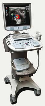
ZONARE?s z.one system allows for real-time consultation at the point of care and features a small footprint.
The Mountain View, Calif-based ultrasound manufacturer recently unveiled a number of new transducers and other products. On the pediatric front, the company released the C9-4t for pediatric head and abdomen imaging. The tightly curved, small-footprint transducer offers up to 134 degrees of imaging with 2D frequency choices from 9 to 4 MHz.
In addition to its small footprint, the cart?s portability made the z.one an advantageous choice for Columbus Children?s Hospital. ?The ability to maneuver the small footprint cart into tight spaces in the intensive care units is key, as we do more and more portable studies on critically ill children,? said Coley. ?We don?t think that we are giving up any diagnostic quality, so there is no compromise. For physical stress on the sonographers, this is also a clear win.?
Coley added that one of the biggest benefits with the z.one has been the ability to deploy images wirelessly over the hospital?s secure LAN, which allows the radiologists to see images as they are being performed, and allows for real-time consultation with the sonographer at the point of care to make sure that the right image is being captured so a correct diagnosis results. ?Getting portable studies over the LAN has been a much bigger deal than I originally thought it would be,? said Coley. ?Radiologist and sonographer satisfaction is way up because of that.?
Another surprise was how readily the sonographers embraced the z.one. ?Usually, when you have a new machine, and one that is different from the others already installed, there is a reluctance to use it,? said Coley. ?Initially, [the sonographers] took it because it was easier to push. Now they take it because it produces better images.?
In addition to the new pediatric transducers, ZONARE also recently presented several work-in-progress features for ultrasound imaging of the heart and liver, using its C4-1 Curved Array transducer. This transducer can offer additional diagnostic information in liver lesion assessment by narrowing down the differential diagnosis, similar to contrast-enhanced CT of liver lesions. The C4-1 is a small footprint, curved array transducer designed, according to the company, to scan technically difficult-to-image patients. The transducer includes improved access and performance, including increased penetration?up to 30 cm?with sensitive Doppler imaging.
The P4-1c cardiac transducer is designed specifically to help image the left ventricular wall and endocardium?also in technically difficult-to-image patients?by opacifying the left ventricle with contrast, making it easier to evaluate wall motion.
The new L14-5w Linear Array transducer is designed to improve the imaging of small body parts, breasts, and superficial anatomy. It has a 55-mm-wide field of view, which can be expanded using the company?s virtual apex array technology.
Several new applications have been added to the z.one. Elastography applications for breast imaging offer a qualitative visual assessment of the mechanical stiffness properties of tissue. The high-resolution images are generated and visualized using a variety of grayscale and colorized maps. The C8-33D enables z.one systems to capture 3D images. This capability is designed primarily for use in obstetrical imaging during the second and third trimesters. The C8-33D is a curved linear transducer that offers mechanical sweep array, 3D fetal surface rendering, 3D multiplanar rendering, and other diagnostic tools. According to the company, the C8-33D may reduce exam time, allowing physicians to spend additional time on patient care.
?C.A. Wolski
Spotlight on Study: Ultrasound and Breast Imaging
While debate and political wrangling about screening mammography echo noisily through the corridors of power in Washington and elsewhere, a study, sponsored by U-Systems, is quietly examining the efficacy of ultrasound as an adjunct to traditional x-ray breast exams.
The study could not have come at a better time, said the Sunnyvale, Calif-based company?s president and CEO, Ron Ho, particularly after the recent recommendations issued by the American College of Radiology and the Society for Breast Imaging, which said ultrasound should be used to screen high-risk women who have a family history of the disease or a mutation of the BRCA1 or BRCA2 genes.

U-Systems? somo-v ABUS offers a more complete view of a dense breast, say experts.
The company?s nationwide study, SOMO-INSIGHT, was begun in early 2009 and will conclude in late 2010. It will include 10 sites in total and 20,000 patients.
Staying on the cutting edge of early breast cancer detection was the primary reason that Florida-based Radiology Regional Center opted to be one of the study sites for the SOMO-INSIGHT study. ?Automated breast ultrasound (ABUS) allows for the standardization of a modality that lends itself as a cost-effective screening tool for women with breasts that are at least 50% dense, in conjunction with screening digital mammography,? said Mary Kay Peterson, MD, director of women?s imaging.
Peterson noted that the ABUS method has some important advantages for women by catching breast cancers earlier. ?More and more premenopausal women are being diagnosed with breast cancer, in part due to improved imaging, increased awareness, better education for caregivers and patients, and better access to health care. However, women who are premenopausal tend to have aggressive breast cancers, therefore, the sooner they are detected, the better the outcome for the patient. This means increased survival and decreased financial costs due to treatment for a less advanced presentation,? she said.
According to Ho, other studies have already proven that ultrasound is advantageous for imaging women with dense breasts (which, he says, accounts for about 40% of the US female population). However, the problem with these earlier studies, he said, is that they used a handheld ultrasound detector, so the images could be viewed only in real time with the radiologist present and not recorded for later review or historical comparison.
U-Systems? somo-v ABUS has eliminated this stumbling block by allowing images to be recorded, stored, and reviewed. The somo-v captures a 3D image with the patient in a supine position, using the company?s linear high-definition ultra broadband transducer. According to Ho, the somo-v can image a breast and the scan can be reviewed in about the same time and in the same way as a traditional x-ray mammogram. The system also includes a pattern recognition system that automatically detects the patient?s anatomical tissue signature and optimizes the system scan parameters, customizing each scan, according to company literature.
For Peterson, the advantage of the somo-v ABUS is a more complete view of a dense breast, in a much more cost-effective way.
The system itself is portable?about the size of a traditional portable ultrasound machine?and can run off a normal wall socket and easily interface with a department?s PACS system. However, the price for the machine is about half that of a traditional x-ray mammogram system, and, Ho says, the return on investment is about 6 months.
Data collected this year will be used in an effort to secure payor reimbursement approval. Data collected in 2009 will be used by U-Systems as part of the Premarket Approval process to secure Food and Drug Administration approval for its somo-v ABUS. Every site taking part in the study is required to meet specific criteria, including patient screening volume, being MQSA certified, and already using digital mammography detectors.
?C.A. Wolski
Toshiba Ultrasound Goes Portable
Riding the wave of a growing demand for ultrasound in specialized departments outside radiology, Toshiba America Medical Systems Inc, of Tustin, Calif, recently received FDA clearance for its Viamo laptop ultrasound system.
Making its US debut during the 2009 Radiological Society of North America meeting in Chicago, the Viamo is described as ?the industry?s no-compromise ultrasound system,? equipped with advanced radiology functions that had been previously unavailable on hand-carried systems. According to Erin Owen, senior product manager for Toshiba?s ultrasound business unit, the company received much positive feedback about the device during the annual meeting. Customers were particularly impressed with how its small size did not compromise image quality, as well as the fact that they could use their existing transducers.

The Viamo laptop ultrasound system includes sophisticated capabilities like Advanced Dynamic Flow.
?The biggest demand is that ultrasound is going to a lot of different areas now,? Owen said. ?In the past, ultrasound really only went to radiology. But now you?ll see it in emergency departments, in surgery, endocrinology, OB/GYN, pediatric, and other niche markets.?
Technology migration is a key benefit of the Viamo, which the company says boasts image quality comparable to that of larger, more expensive ultrasound systems, but at a more affordable price point given its small size. ?Many features we migrated from our high-end system, like Advanced Dynamic Flow, which lets us see smaller vessels better with color,? Owen said.
Owen noted that the Viamo was not developed as a replacement for larger cart-based systems, but rather as a bedside supplement to them. For example, the laptop ultrasound can be used during liver transplants when medical personnel must image the anastomoses to assess blood flow through the vessels. ?The Viamo is specifically designed to provide advanced radiology capabilities in a portable system, creating more comfortable exams for patients,? said Girish Hagan, Toshiba?s vice president of marketing. ?Providing the best value in the hand-carried segment, the Viamo delivers high-quality images for numerous clinical applications.?
With the Viamo, current Toshiba customers can interchange their transducers while using the Viamo?s transportation pole. The company says this flexibility will allow hospitals to ease health care costs by reducing the need to purchase multiple transducers. New customers will be able to use Viamo transducers with other Toshiba ultrasound systems they may acquire in the future. The product was also designed for customer ease of use, carrying a simple touch-screen interface that is programmable in panel or tablet modes.
?The Viamo provides the best value in the hand-carried class by offering a feature-rich, portable system at a reasonable price point,? Hagan continued. ?With the ability to perform high-end radiology exams using a portable laptop system, Viamo rounds out Toshiba?s ultrasound product line, delivering function and value in the hand-carried segment, along with the exceptional image quality and clinical accuracy Toshiba is known for.?
Toshiba is ready to begin shipments right away and is now taking orders for the recently cleared technology. The company began delivering systems for its demo sites in mid-February.
?Elaine Sanchez


