MRI is considered a very sensitive modality available for the detection of primary or recurrent breast cancer and has proven to be a valuable tool for the detection and characterization of breast disease, assessment of local extent of disease, evaluation of treatment response, and guidance for biopsy and localization.1 The American Cancer Society recommends patients with a 20% or higher lifetime risk for developing cancer start screening at age 30, and receive both screening mammography and breast MRI annually.2 For women with dense breast tissue, breast MRI is able to better detect certain hard to spot breast cancer lesions missed by mammography. However, a particular challenge for interpretation of breast MRI images is the assessment of a lesion’s morphologic and kinetic features, which can be time-consuming.

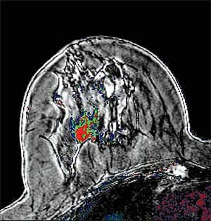
SpectraLook assists radiologists in reviewing large MRI datasets by creating colorized images based on signal changes defined by tumor physiology. As a result, radiologists receive qualitative and quantitative information about the breast tissue using colorized maps that highlight areas of suspicion. COURTESY OF ICAD
To address some of these challenges, image analysis technology has begun to play an increasingly important role in breast cancer detection. Compared to standard mammography, breast MRI with advanced image analysis provides radiologists with a more detailed and accurate visualization of the scan to help better determine the size and location of tumors. Given its ability to find more or fewer lesions than initially found with mammography or MRI alone, it also can serve as a guide during biopsy or other treatment.
SpectraLook® from iCAD provides objective, consistent, quantitative analysis for cancer detection, localization, staging, treatment planning, and serial monitoring. SpectraLook assists radiologists in reviewing large MRI datasets by creating colorized images based on signal changes defined by tumor physiology. As a result, radiologists receive qualitative and quantitative information about the breast tissue using colorized maps that highlight areas of suspicion. This image analysis tool is of significant benefit in situations where the morphology analysis is ambiguous and additional information is needed to make a clinical decision.
When coupled with VersaVue® Enterprise, iCAD’s image review and analysis software, SpectraLook offers a comprehensive solution that improves the analysis, interventional planning, and reporting of breast MRI results while streamlining the radiologist’s workflow and diagnosis. An advanced pharmacokinetic model assesses changes in signal intensity to calculate numerical values of key physiological parameters. This allows clinicians to discern specific biological processes occurring in malignant versus benign tumors. iCAD’s analysis software has been shown to both reduce potential errors and variation associated with qualitative analysis and improve consistency across magnets, image acquisition protocols, and clinicians.
iCAD’s PrecisionPoint® software is an integrated module within VersaVue Enterprise, which provides radiologists with an automatic calculation of the location and depth of a targeted region of interest, making MRI-guided breast biopsies easier, faster, and more reliable.
In my own group practice, iCAD’s SpectraLook software has allowed each of the radiologists to have their own specialized and individualized breast MRI hanging protocols when reviewing the vast accumulation of data from all of the images obtained from each breast MRI case. This particularly satisfies each of the radiologists who interpret and read each case and customize it to their specifications. SpectraLook software also allows each radiologist to easily cross correlate multiplanar imaging and to process the images from multiple scanners that perform either axial or sagittal breast MRI imaging. Since the radiologists in my group read from multiple sites (which include a hospital and multiple outpatient offices), SpectraLook software allows universal viewing and interpretation of the cases from multiple facility locations. SpectraLook software has accommodated the continued growth of increasing numbers of breast MRIs in my practice and has provided an extremely useful method of breast MRI interpretation, meeting the individual needs of the various radiologists in my practice.
Grace Malantic-Lu, MD, is chief of Breast Imaging, Edison Radiology Group, at JFK Breast Center in Edison, NJ.
REFERENCES
- ACR Practice Guideline: Performance of Contrast-Enhanced Magnetic Resonance Imaging (MRI) of the Breast 2008, p 561
- www.cancer.org/Cancer/news/News/acs-advises-mris-for-some-at-high-risk-of-breast-cancer