Digital radiography systems convert X-ray signals into digital images at the point of acquisition, thus minimizing artifacts and distortions that are often associated with conventional radiography systems. Incorporating computer technology for capturing, displaying, enhancing, and storing images, digital methods provide obvious advantages over film. Contrast value and image brightness can be modified to optimize visualization. Also, images can be altered to meet clinical requirements.
Patient records, exposure settings, image review, distribution, and archiving are all done automatically when DR systems are combined with a picture archiving and communications system (PACS) and/or a radiology information system (RIS). DR provides yet another means to increase productivity.
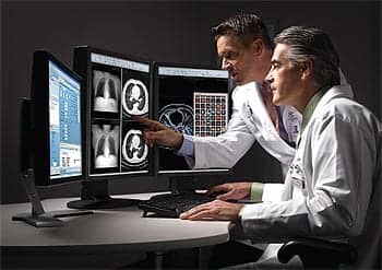
In a cardiac catheterization lab, DR helps doctors obtain the information they need to optimize patient care. Quality of images and the ability to manipulate them through the use of DR also helps physicians determine the best place to insert a drug-eluding stent to prevent coronary restenosis. DR optimizes many other cardiac cath lab procedures, in turn helping improve the lives of many patients.
Frost and Sullivan industry data estimated that nearly 4.5 million cath lab procedures were performed in the United States in 2000. Many of these procedures were optimized through the use of a DR system.
Digital Network Initiated in Cleveland
In 1991, physicians from the Cleveland Clinic approached Philips Medical Systems with the idea of making a digital network. But the technology was just not there, according to Frederick Heupler, Jr, MD, director of the diagnostic section of the Sones Cardiac Catheterization Lab at the Cleveland Clinic Foundation (CCF of Cleveland). CCF staff was persistent, however. In both 1995 and 1996, physicians interviewed 13 cardiac cath lab vendors, again with the idea of developing a digital network. None of the vendors made products that met CCF’s very detailed specifications for the performance of a digital network, so the clinic collaborated with Philips to build one—with specifications totaling 30 pages. CCF doctors performed all of the testing themselves in order to determine what specifications were best for the type of system they wanted.
The doctors didn’t just want the digital system for CCF. They wanted Philips to include the system in a product line so that it would be available to other hospitals and medical centers that wanted to convert their cardiac cath labs to digital systems. Today, the CCF is film free and for the past 4 years, it has relied completely on digital technology. “Our intent was to meet every advantage of a film distribution system plus the added advantages of digital imaging,” Heupler says.
One of CCF’s specifications was an open architecture so that the system could interface with other vendors’ products at other hospitals and exchange information easily. Doctors at CCF did much of the testing themselves to determine the best image quality and resolution. Angiographers scored different types of image formats for spatial resolution (to detect small pieces of calcium in a coronary artery); certain aspects of contrast resolution (overlying spine or diaphragm and heart-lung interface); temporal resolution (to check for any lag or blurring); and brightness (the doctors’ perception of the luminescence of the monitor).
“We had to decide ourselves what would give us adequate image quality,” Heupler explains. “We evaluated film versus digital and different degrees of image compression. We compared color monitors to black-and-white monitors for image quality. And we compared 512-pixel to 1,024-pixel monitors.”
Another specification required each review station to cost a maximum of $5,000—rather than the $35,000 apiece that was quoted to CCF by different vendors.
“With 50 review stations to replace, even the Cleveland Clinic couldn’t afford to pay $35,000 each for them,” Heupler says. “We needed 50 review stations, complete with a viewer and controls, so that the user could sit down with virtually no instructions and use it. It had to be very user friendly. The monitor had to be the same as the others we had. We also wanted to have access to images done within the past month, and we wanted the archive to be accessible within 5 minutes, with storage on a local file server.”
CCF also tested the reliability of various data storage systems. Prior to the digital transition, 3 employees were responsible for the film library. Despite their best efforts, about 5 out of 10,000 films were lost each year. “We didn’t want to lose any more than that,” Heupler says.
CCF Physicist Robert Cecil, PhD, collaborated with Philips to design the clinic’s digital storage system. His concept was that every major institution should have a digital information warehouse, because all information will eventually be stored in digital format. CCF’s digital storage warehouse has a capacity today of 3,000 terabytes. The equipment that houses the storage system is about 7 feet high and 15 feet wide.
The system itself is controlled robotically with a digital tape storage system. A robotic arm selects the tape that contains the requested image, downloads it, then removes the tape and puts it away when the user is finished. The entire process takes just a minute or two—dramatically faster than tracking down a film canister.
“We wanted to be able to distribute images between cath labs. That way, if a doctor calls me from one of our outlying cath labs and wants me to review a case to see if the patient should have an angioplasty, for example, I can access it in a minute and a half,” Heupler says. “We wanted to have a very high reliability of storage information.”
In the end, Philips helped CCF reach its goal of a digital network, meeting all of CCF’s specifications. Heupler adds, “Their compliance was a key part of the whole project.” Although the process took about 6 years, the bulk of that time—about 90%—was spent developing and designing the system. “Now if someone wanted to transition a cardiac cath lab, it can be done much sooner,” he says, “in a matter of months.”
Flat Panels Leading the Way
“Flat-panel detectors, which are a combination of cesium iodide and amorphous silicon detector matrices, are available for use in cardiac catheterization visualization,” says Gregg A. Cohen, PhD, principal consultant with Xtria Digital Imaging Solutions (Dallas). “These detector matrices are being used in place of optical image intensifier systems.”
Despite differing opinions, Cohen believes that in the purest sense, true DR is not being used in cardiac cath labs. “Typic-ally, DR uses a direct access amorphous silicon or amorphous selenium matrix with a read-back time on the order of 5 seconds. The spatial resolution of these amorphous silicon panels is on the order of one tenth of a millimeter. Due to these spatial and temporal constraints, it is generally inappropriate for interventional vascular visualization,” he notes.
The charge-coupled device (CCD) or chip that has been instrumental in television technology has made it possible for flat-panel detectors to provide such detailed and high-quality images, according to Marcia Wroblewski, field marketing manager for Philips Medical Systems (Bothell, Wash).
“The whole efficiency of technology leads to better imaging and better diagnostics,” says Wroblewski, who believes that DR is being used in the cardiac cath lab—and quite a bit. “About 75% of cardiac cath labs now are using DR. It opens up the opportunities to share information across the spectrum of healthcare.”
Heupler concurs, adding, “I review 400 to 500 angiograms per year. About 80% to 90% of the images I receive from outside institutions are digitally acquired.”
Some of the technologies that make DR possible were not available for use in radiology until the past few years. Manufacturing advances and growing general acceptance of flat-panel technology have enabled progress in this area. The impetus for this change lies in manufacturing advances that have made significant improvements in spatial and temporal resolution as well as general reliability, according to Cohen. Computational power has increased while prices have decreased, making both the software and hardware support more affordable.
Advantages Over CR
William A. Gray, MD, is an interventional cardiologist and director of endovascular care for the Swedish Heart Institute at the Swedish Medical Center (Seattle). Gray cites many advantages of DR, not just in his cath lab, but in cath labs overall.
“The quality of the image is excellent. We can modify the quality of the image, play with the width and height, and adjust the grayscale. We can modify the pixels and edit the image,” he says. “DR means less repeated images, because we can see what we need, especially in peripheral vascular imaging. DR adds significant mobility to imaging, with the utilization of PACS, image transfer, and image manipulation. It’s a major advantage over conventional radiography.”
Research from Canon Research Center America1 (Palo Alto, Calif) shows that DR without RIS results in substantial workflow savings over traditional film/screen practice. An additional 30% is reduced in total exam time using DR with RIS integration.
“We don’t have a film developing room, so we don’t have to worry about chemicals and developing images,” Gray explains. “Archiving is much easier with DR. The images go onto CDs, which are much smaller in volume than a can of film. The space required to store the same number of cases is much less. The CD can be played on virtually any PC. The transfer of images is much easier than with film. We can copy all digital studies much easier. Although we don’t have PACS, DR allows system-wide access to the image.”
Cohen adds that the main benefit to using flat-panel technology is the ability to capture true digital data, and that calibration is simple and reproducible.
“Additionally, the lower exposure to radiation necessary to generate an image is a significant advantage, driving adoption of this technology,” he says. “The utilization of this technology, however, is often driven by economic factors. Adoption of flat-panel technology requires a technology foundation that is based on digital PACS for the capture and storage of digital images. Often, financial costs associated with the replacement of multiple rooms are the driving factor, rather than technology limitations. An average cardiac invasive laboratory replacement with imaging equipment is often in the range of a $1 million capital expense.”
According to Cohen, facilities that choose to invest in new DR technology—if it is a new construction project—are more likely to have multiple labs as well as a PACS to support the capture and archiving of all cardiac images. If it is a replacement room for cardiac, it is more costly to move directly to a completely digital environment and a single PACS. Frequently, cost constraints force the organization to operate two systems to capture, manage, and archive images, he says.
Wroblewski agrees, adding that physicians and hospitals both benefit from the use of DR in cath labs. Physicians are able to share information with other physicians in different geographic areas, whether in a particular case or an entire study. Hospitals can realize a significant financial benefit from utilizing DR, not only in their cardiac cath lab, but also throughout the hospital and/or system.
“Multimodality imaging offers the opportunity to coordinate exams and/or to collaborate with each other, and it plays a role in diagnosis and treatment. DR helps the cardiac cath lab become more efficient,” she explains. “The amount of detail in an image is critical in helping cardiologists place catheter balloons in tiny coronary vessels.”
Doctors at CCF initially thought that the spatial resolution of film was higher because the resolution of a grain of film is much higher than that of a digital image. However, they later realized that contrast resolution is much more important than spatial resolution.
“The contrast resolution in film is very poor. There’s a lot of black and a lot of white, but very little gray. With digital imaging, you can adjust the relative intensity of each pixel. You can adjust and control the grayscale,” CCF’s Heupler explains. “The contrast resolution is much better with digital. We wonder now how we ever used film. We used to think that film was the gold standard, but it is far inferior to digital.”
Another advantage of digital imaging is that a digital copy made of a digital image will be the exact same thing. In other words, the quality will be the same, with no distortions or change in resolution, Heupler notes.
“Digital has totally changed the way we do things. We don’t need to wait 20 minutes for film to be developed. We used to wait a minimum of 20 minutes to develop film, and the film was usually batched, so sometimes we had a 2- or 3-hour wait to get an image to review. Now we can review images immediately after [a procedure],” he says. “You can have information provided to the person who’s taking care of the patient much more quickly [with digital imaging]. If I do an image for a noncatheterization cardiologist, they can have the image within minutes to review with the patient or a surgeon. We don’t need to carry film around from one place to another.” For example, if Heupler finds something rare or unusual when performing a catheterization and he wants another doctor’s opinion, the image can be pulled up and looked at right away.
“And if you want to use images for educational purposes,” he says, “you can distribute the image at a conference and project it onto a screen for everyone to see.”
Initial Resistance Easily Overcome
CCF was ready to install its new digital system in late 1997. Many of the physicians at the clinic had been using film-based systems for 10 or even 20 years, and some were reluctant to give it up. One physician was adamant about not using a computer.
“We installed the new digital review stations only in the offices of those doctors who wanted it,” Heupler recalls. “We still sent them film and stored the digital images for them to review. A doctor in one office might have had a digital review station, while the doctor in the next office didn’t. It took 6 months for everyone to see the advantages of the digital system, and then everyone wanted digital. No one wanted to receive the film anymore. Even the doctor who had so clearly resisted said that he wanted a digital review station—and he wanted it by the next day.”
Clearly, those who have been exposed to digital imaging—especially in cardiac cath labs, where digital can be crucial in diagnosing the best course of action for a patient—can’t imagine ever going back to film. The possibilities for digital imaging are exciting and almost endless. Being able to manipulate and fine-tune images in order to focus on a particular procedure or element has opened up many opportunities for cath labs to optimize patient care.
Laura Gater is a contributing writer for Medical Imaging.
Reference:
1. May GA, Deer DD, Dackiewicz D. Impact of digital radiography on clinical workflow. J Digit Imaging. 2000;13,2(1):76-78.