 |
I remember how excited I was to start using digital mammography. During my fellowship in 1998-1999, digital mammography was in its infancy, but the Hospital of the University of Pennsylvania was involved in the initial research studies that validated digital mammography as being just as sensitive as the gold standard, the tried-and-true analog film-screen mammogram. My first exposure to digital mammography quickly showed me how many more calcifications I could see and how suddenly dense breast tissue seemed less dense. Being a fellowship-trained breast imager, I have always prided myself in trying to stay on the cutting edge of my field. I was excited to be one of the first radiologists in New Jersey to be certified to read digital mammography early in 2001. I remember flying to Chicago (which was one of the only places to go at the time) to take my required 8-hour course to obtain my certification. What I didn’t realize during the course was the learning curve that would be required to transition from reading film screen to reading digital studies. Now, initial digital training can be performed at your own facility without having to go to an outside course.
During the first year of reading digital studies, we were required to evaluate the studies on printed hard-copy films because soft-copy reading was not yet FDA approved. On the one hand, if you just read digital mammography on hard-copy images, your transition to digital mammography would seem quite easy, as your reading protocols would not change much. However, you would lose many of the added benefits of reading digital mammography such as the ability to change window/level to maximize visibility as well as the ability to have significantly improved magnification. On the other hand, reading both hard and soft copy significantly increased the amount of time necessary to read the studies. Thankfully, for those who are transitioning now, you can go straight to soft copy, which is much better. The dual reading, however, was even more labor intensive at the time because our viewing setup was far from ergonomic. Our mammo alternator was at least 3 feet away from our soft-copy workstation, and our mammography reading room at the time did not make for the most conducive transition to digital mammography as the room was built long before the invention of digital mammography. Also, the digital workstation viewing protocols were much less sophisticated than they are today, so it took much longer to perform the soft-copy evaluation.
The First Challenge: More Findings
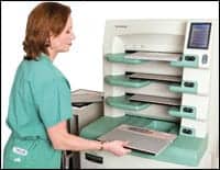 |
| A multiplate digital mammography image reader like this one from Fujifilm is ideal for centralized locations. |
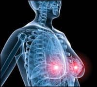
Probably the most challenging aspect of my transition from film screen to digital mammography was trying to decide what to do with all the calcifications I could now see. Everyone I know, including myself, had a significant initial bump in her/his recall percentage rate particularly relating to calcifications. I have had colleagues tell me over the years during their transition to digital that they didn’t like it mostly because of an “ignorance is bliss” philosophy. Seeing more calcifications, which statistically are more often benign than cancerous, can be very problematic. Nobody wanted to add to patient anxiety to evaluate a finding that the patient had likely already had for years. Yet how you could you ignore what seemingly appeared to be a new finding? The management difficulty still continues even now, years later, as I still have many patients who come from outside facilities where their prior studies were analog. With time and experience, my colleagues and I have learned better which calcifications need further workup and which calcifications can just be left alone.
On a more positive note, a lot of the previous technical callbacks have decreased significantly when I transitioned to digital. But most importantly, the DMIST trial in 2005 demonstrated that digital mammography is more sensitive than film-screen mammography in women who are premenopausal or perimenopausal, as well as women of all ages who have dense breasts. On a personal note, having years of experience with both technologies, I have no doubt in the results of the study. Dense breasts are significantly easier for me to read digitally than they ever were on film screen. Although the research didn’t show any added benefit, I personally find digital mammograms in women with fatty breasts or those with scattered densities also to be easier to read. Visualization of breast tissue in patients with implants is also less challenging than with analog studies.
Setting Up the Reading Room
A big part of the digital transition is the initial setup of the reading space. This should be well thought out and not done haphazardly, because once set up it is not easily undone. The initial financial investment in room development, including ergonomic desks and chairs as well as appropriate masking of surrounding light, is well worth it. If multiple radiologists will be sharing a workstation, one should consider buying one of the many available adjustable desks to accommodate the different heights of the physicians. I have seen many radiologists have new neck and wrist strain during their digital transition.
One of the difficult challenges of transitioning to digital mammography is how to handle the first few years when the prior mammograms are all analog but the current study is digital. Reading soft copy for the digital mammogram is clearly advantageous and allows the radiologist to use all the available tools to manipulate the images, so I would not recommended just printing out the images and reading the hard copy. By doing this you would also negate the cost savings of not using film in the digital environment. The most widely used transition setup is having the prior studies hung on an alternator at a shallow angle to or above the soft-copy workstation. Another option, if you have the available manpower, is to digitize a more recent as well as an older prior mammogram and have it sent back to the soft-copy workstation for a more direct comparison. Having personally used both options, I prefer to have the priors digitized because it allows for significantly faster reading and, in my opinion, significantly less neck strain. Another added benefit is not having to constantly turn off and on the lights from the alternator, which is time-consuming and cumbersome.
Demand for Digital
 |
| While many still consider FFDM the breast imaging gold standard, CR mammography is also a good option for those who want to add digital mammography to their practices. |
Another obstacle to tackle in the digital transition it how to handle the situation when your facility has both film-screen and digital units being used concurrently. It is preferable to be in this situation for as short a time as possible because many issues arise quickly. The first issue is how do you handle scheduling? Ideally, you would want all women with dense breasts as well as the premenopausal and peri-menopausal women to have a digital study, but it is very difficult to schedule patients in a high-volume facility with this in mind. Also, patients in general are informed consumers and are very aware of the benefits of digital mammography, so they will start requesting it once they know your facility has it. This leads to long waits for the digital room and the film-screen room being underutilized. The waiting time for the patients will go up significantly, which will lead to more anxiety in the waiting rooms—the last thing that a breast center wants or needs.
The radiologist also will have a harder time in this process because she/he will be constantly switching brain gears between reading film-screen and digital studies. One word of advice: For any women who are “callbacks” from a screening study, the extra diagnostic views should be performed using the same technology as the initial screening study. It is most difficult and potentially unnerving to read a digital-screen mammogram but then do extra views using an analog device, particularly if the potentially abnormal finding such as calcification is not seen on the additional diagnostic views.
With all the challenges that I mention here, I am thrilled to only be reading digital mammograms at Montclair Breast Center. In fact, when I have to do outside consultations on film-screen studies, I often feel amazed at how much more I can see using the digital technology. Frequently on extra digital views performed during the consultation, particularly on women with breast cancer, I am somewhat shocked by the improved visualization and the unanticipated larger extent of disease. Occasionally, I end up repeating the full four-view mammogram for the patient. I do not know anyone past his/her learning curve for digital mammography who would want to return to film-screen reading. In fact, most of us believe that our speed in reading has even improved enough with digital to not seem like as much of a difference from analog as it used to.
 |
Future technologies that build on digital mammography, including digital tomosynthesis, are also just around the corner, as displayed most impressively at this past RSNA meeting. My hope is that as the years go by the cost of converting to digital will go down so that it will be practical even for the smallest of breast centers. The difficult transition to digital mammography is well worth it, and I have no doubt that it is only a matter of time before film-screen mammography will no longer exist.
Rose Heller-Savoy, MD, is the director of mammography services at Montclair Breast Center, Montclair, NJ.
