 |
· Images in Your Pocket
· TWO ARE BETTER THAN ONE: Sectra Showcases Integrated RIS/PACS
· TEAMING UP ON TECHNOLOGY: Freedom to Travel
· Premier and Philips Partner on PACS and More
Images in Your Pocket
Walk, text, e-mail, rock out to iTunes … review diagnostic images?
The Apple iPhone 3G, released in July, is a device that aims to meet the everyday needs of its customers, which include clinicians. So the company has launched its iconic phone with add-on applications that offer medical imaging capabilities.
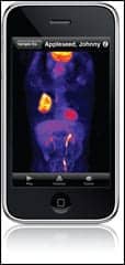 |
| The MIM application for the iPhone allows for multimodality volumetric imaging with fusion. |
One of these applications is the MIM for iPhone, provided by MIMVista Corp, Cleveland.
“The MIM application for the iPhone is the essence of cool for a radiologist who thrives on image display,” said Peter Faulhaber, MD, director, clinical PET, at University Hospitals Case Medical Center in Cleveland.
MIMVista’s Chief Technology Officer Mark Cain explained that the software allows for multimodality volumetric imaging with fusion. It provides multiplanar reconstruction of data sets from the range of modalities, including CT, PET, MRI, and SPECT. Furthermore, users can change image sets and planes and adjust zoom, fusion blending, and window/level, using the phone’s multitouch interface.
The software had been around for the past 5 years, but for use with a MIM workstation, Cain said. When Apple released the beta version of its software development kit, MIMVista signed up and ultimately decided to build a mobile version of its existing software. This version, the application for the iPhone, is meant for consultative purposes, not diagnostic.
“The iPhone application is primarily a remote viewer,” Cain said. “Radiologists are tied to their workstations, and sometimes they just need a way to see the images when they’re not at the workstation. But this is not intended to be a diagnostic workstation in your pocket.”
Cain said the application is also useful for referring physicians, who will not need to set up medical imaging viewing workstations for patients to look at their images. “That’s expensive and cumbersome for a lot of sites,” he said. “Now the referring physician can literally carry the images in his pocket, sit down with the patient wherever the patient is, and show him or her what the radiologist saw.”
Cain said there is obviously a difference between a $20,000 radiologist monitor and the iPhone. However, the mobile device “has a fantastic display.” In fact, the brightness is even better than some monitors, he added. While users won’t be able to see many images at once due to space constraints, physicians seem to be in agreement that it is very good for the purpose it serves, Cain continued. Apparently, Apple felt the same—it honored MIMVista with a design award.
“The software is fast and intuitive,” Faulhaber said. “I think referring physicians will be able to seamlessly review a patient’s images while consulting over the phone. Patients will be even more impressed.”
All communications are transmitted over a secure connection that uses password locks, tamper prevention, and data encryption to ensure patient privacy. Individual patient data can be downloaded to an iPhone or an iPhone touch from a MIM Workstation or MIM Storage Server.
The application is free from Apple’s App Store at www.itunes.com/appstore under the Healthcare and Fitness category. Also available is the other medical application for the iPhone, called Modality, which features illustrations and labels of the human anatomy and functions as a reference guide for medical students.
MIMVista is now working on a professional version that looks to provide the features that radiologists would need on a regular basis, Cain said. “They want to use it more, they want to accomplish more, they want to do more measurements,” he added. This will be accomplished in the later version.
—Elaine Sanchez
Two Are Better Than One
Sectra Showcases Integrated RIS/PACS
PACS is critical to the management of images while a RIS is key to managing workflow. Together, the two can provide a department, facility, or entire health care organization the ability to more effectively and efficiently manage its imaging business. Subsequently, many institutions have sought or are seeking the integration of these systems, along with other electronic solutions, such as the EMR, billing records, and practice-management software.
One newer option on the market today is the integrated RIS/PACS solution from Sectra Medical Systems, Shelton, Conn. The company displayed the solution, available for the US and Canadian markets, at the AHRA [American Health Radiology Administrators] 2008 Annual Meeting held this past July in Denver.
Working together, integrated RIS/PACS systems enable administrators to leverage strengths and identify weaknesses of the organization as a whole, improving productivity and patient care. When data is shared, it eliminates manual intervention, reducing errors, improving information collection, and increasing productivity.
Easy exchange of data can improve billing accuracy (and sometimes revenues), while documentation can help with future diagnosis, prognosis, and treatment as well as regulatory and legal requirements.
Workflow is smoother with prepopulation of data, worklists, and associated materials, such as images. Any relevant information is at the physician’s fingertips, meaning clinical decisions may be better informed. Treatment progress may be more comprehensively monitored through previous reports, prior images, and treatment protocols that have been recorded and are readily available through an integrated system.
Many PACS solutions are able to integrate with other vendors, and Sectra’s PACS is no exception. Sectra has achieved various levels of integration with all of the leading RIS solutions, a range that includes worklists, requests and reports, status information, prefetch, patient demographics updates, data integrity checks, and desktop integration with the display software. With its own PACS, the Sectra RIS uses brokerless integration.
Sectra’s PACS is built on a Web-based foundation with features, such as zero client administrations, roaming desktop, and concurrent users, that allow it to be easily deployed over an entire enterprise. Teleradiology is easily enabled wherever the physician is located.
The system is scalable and designed to work efficiently over high-latency networks; Sectra’s PACS has demonstrated uptimes of 99.99%, according to the company.
A variety of workstation options (such as a paper-free radiology workstation and a dedicated mammography review workstation) and add-ons (such as 3D, cardiology, and nuclear medicine packages) are available.
The Sectra RIS solution is designed for a 100% film-free and paper-free environment. Created to improve radiology workflow, features permit efficient planning, optimized throughput, comprehensive documentation, thorough follow up, and accurate billing.
Data mining and report capabilities expand with system integration. Administrators can use the collective data to determine the most profitable modalities, the most active referring physicians, employee productivity levels, the busiest time periods, and patient throughput rates.
Sectra’s integrated RIS/PACS solution can help customers who have multiple sites or providers. Austin, Tex-based Solis Women’s Health leverages Sectra’s RIS/PACS technology at multiple breast imaging centers. The organization has sites in Arizona, North Carolina, and Texas.
“Our organization requires maximum operational flexibility for our growing network of centers to be able to realize the full potential of our business strategy,” says Brad Hummel, CEO of Solis, in a Sectra press release. “Sectra meets this requirement with an integrated RIS/PACS solution that is scalable, reliable, easy to implement, cost-effective, and robust.”
—Renee DiIulio
Teaming Up On Technology
Freedom to Travel
Pairing a mammography image viewer with a media writer optimizes the portability of breast images.
The American Cancer Society recommends that women age 40 and older have an annual mammogram to screen for breast cancer. Early detection can lead to better outcomes, particularly if caught before the disease has spread to other regions of the body. Comparison to previous exams is a key part of this screening effort. Yet patients will not necessarily return to the same physician, year after year. Families move, physicians move, and stuff happens—many women find themselves lugging films among providers over the years.
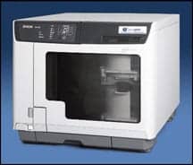 |
| PACSGEAR’s MediaWriter disc burner is now compatible with Three Palm’s MammoViewer mammography DICOM viewer. |
This solution is risky, in terms of losing film, and costly, in terms of printing film. But as the medical community slowly converts to an all-digital environment, so, too, do these films. A new partnership between PACSGEAR Inc, Pleasanton, Calif, and Three Palm Software LLC, Los Gatos, Calif, aims to accelerate that conversion. PACSGEAR’s MediaWriter disk burner is now compatible with Three Palm’s MammoViewer mammography DICOM viewer. Women and physicians can now benefit from a portable mammography file.
“You can digitize the mammography priors—if they are on film—put them with the current study, write the report, and store it all on a disk,” said K. Thomas Pickard, vice president of marketing and business development at PACSGear.
The Three Palms MammoViewer is a mammography DICOM viewer that permits users to display and manipulate a mammography study anywhere. It can be included on CDs, DVDs, and other portable media to enable viewing of the mammography files.
The device displays MG images from multiple vendors, CAD structure reports (shown as an overlay on the image, which can be turned on and off), and digitized prior mammogram film images. Viewing features include multiple image layouts, image-manipulation tools (such as window level, zoom, pan, and insert), and image markup and measurement.
The MediaWriter by PACSGEAR states its function within its name: to write DICOM-structured studies and reports to media, including CDs, DVDs, memory sticks, and flash memory cards. The software/hardware solution features an integrated viewer and a CD/DVD burner with a built-in printer that automates media labeling.
The system is scalable and able to meet the needs of image-intensive departments, including radiology, cardiology, gastroenterology, dermatology, and pathology. The company offers multi-CD and single-CD models. CDs are printed within 3 to 4 minutes.
Partnering MammoViewer with MediaWriter addresses the particular needs of mammography files; both portability needs as well as imaging demands. “Mammography studies have some special needs, and the Three Palm software viewer [MammoViewer] lets physicians look at those images easily,” Pickard said. At the same time, the MediaWriter creates standards-based CDs and DVDs using the IHE Mammography Image Profile.
Portability is the primary value of the partnership. “What this means for customers is that, when their MammoViewer is combined with the MediaWriter, they can create portable mammography records,” Pickard said.
The savings of digital media use in film and printing costs can be significant. “It reduces the need to print film each time a mammography is taken, so there is definitely a cost reduction,” Pickard said.
For the hospital customer, this means everything on one patient can be stored easily in one place. For the physician user, reports, images, and overlays (including CAD markers) are easy to access and read. For patients, health care data is more convenient to transport; their entire mammography history can be stored in one spot. “It gives women their complete record to take from place to place,” Pickard said.
—Renee DiIulio
Premier and Philips Partner on PACS and More
A new 33-month contract will give the hospitals and health care sites of the Premier health care alliance, headquartered in San Diego, access to cardiology and radiology PACS solutions from the health care informatics business of Philips Healthcare, a division of Royal Philips Electronics, based in Andover, Mass. Premier’s more than 2,000 member hospitals and 53,000 additional health care sites now have access to Philips iSite PACS for Radiology and the latest release of the Philips Image and Information Management System for Xcelera PACS for Cardiology, Xcelera 2.2.
The contract was signed between Royal Philips Electronics and Premier Purchasing Partners, Premier’s group purchasing unit, and became effective in April. “We’re very pleased that Philips worked with Premier to create an opportunity that affords even hospitals with limited individual negotiating power the ability to acquire advanced technology,” said Merle Peterson, director of medical imaging for the Kettering Medical Center in Kettering, Ohio, and a Premier alliance and Imaging Committee member.
Potential products and vendors were compared on a number of criteria, including overall functionality, multimodality imaging capabilities, service and support, breadth and depth of products, integration, corporate stability, life cycle, and costs. A team comprised of members of Premier’s IT Services, Cardiology, and Imaging Committees evaluated the suppliers in the Enterprise Image Management Solutions (EIMS) marketplace in 2007 and arrived at Philips.
The Products
The agreement expands previous contracts between the two organizations encompassing CT, MRI, molecular imaging, nuclear medicine, PET and PET CT, RAD, RF, surgery, cardiovascular, ultrasound, and patient monitoring systems.
The Philips iSite PACS solution is an enterprisewide, Web-based image-distribution system designed to integrate into an institution’s existing infrastructure. Features include radiologist worklists, patient-ward specific worklists, modality exam schedules, and full views of patients for referring physicians. EMRs can be image-enabled through the system’s advanced API functionality. Advanced radiology reading stations and always online, long-term storage expand functionality.
Philips Xcelera also offers features that improve workflow in the cardiology department through integration. The multimodality image management, analysis, and reporting solution provides one access point to images and data from key cardiology subspecialties, such as CV x-ray, echocardiography, CT, MRI, nuclear medicine, ECGs, and intracardiac (EP) signals. Users have access to an extensive range of integrated, advanced quantification applications on a common workspace and can quickly identify and review all patient studies performed across their patients’ continuum of care. Reports are easily generated using the data-mining tool and, when combined with Webforum, all information can be viewed throughout the enterprise over the Internet.
The systems are flexible enough to provide solutions for the various facilities within the Premier system. “Whether desiring to automate just a single specialty department or looking to create a comprehensive, multi-modality image and information management environment in cardiology, Premier members will find Xcelera to be flexible, scalable, and cost-effective in addressing their cardiology informatics needs,” said Harry Comerchero, national director of strategic accounts for Philips Healthcare’s Cardiology Informatics group.
The Deal
For their radiology departments, customers can choose from a “fee-per-study” or “traditional capital” payment when making their buying decision. Interested institutions can try out the iSite PACS with no obligation through the company’s Pilot Program. This affords facilities the opportunity to judge performance and estimate return on investment before signing any agreements.
When selecting Philips, buyers can expect 24/7/365 service support, performance uptime guarantees, server and software obsolescence protection, unlimited software licenses, disaster recovery, and other benefits at no additional cost, according to Luke Wigger, director of strategic accounts for Philips Healthcare Informatics-Radiology.
Philips expects many Premier members to take advantage of the new contract offering, “whether they are purchasing their first radiology PACS systems or replacing/upgrading their existing systems. In either case, Philips looks forward to working with the Premier membership, assisting them in providing their patients with high-quality, cost-effective health care,” Wigger said.
—R. DiIulio