 |
· Team Imaging
· PRODUCT DEBUT Exam Impact: During and After
Team Imaging
Athletes call the shots on the playing field, but in the physician’s office, the sports medicine specialist dominates. According to the American College of Sports Medicine (ACSM), primary care sports medicine was initiated in the 1970s with fellowships developed in the 1980s and a certification program by the American Board of Family Physicians (ABFM) launched in the early 1990s.
 |
Despite the structured programs, however, these certified physicians vary widely in their study and practice. A survey conducted by ABFM revealed that physicians with a Certificate of Added Qualification (CAQ) in sports medicine hold either MD or DO degrees and vary their practices significantly with location and type of practice. Imaging is a large part of that practice, and sports medicine specialists have their choice of modalities—all are regularly used to image these patients, and they include ultrasound, radiography, MRI, and CT.
At Orthopaedic Associates of Michigan, headquartered in Grand Rapids, about 70% of the patients are x-rayed, estimates Judy Zane, RT, the institution’s director of medical imaging. Zane’s analysis of May caseloads indicates that two of the health care organization’s locations saw 6,000 patients in 1 month. Staff includes 34 physicians, three who specialize specifically in sports medicine and two with other specialties combined with a sports medicine focus.
Ultrasound exams are referred to the hospital; radiography is supported on-site. The main location has two DR machines and one CR; the satellite office is home to one more DR and one more CR unit each. This inventory has grown with the practice with recent acquisitions made to improve patient volume management. “Our downtown facility was landlocked, and patients were waiting 45 minutes to get an x-ray,” Zane said. The additional equipment alleviated the wait.
Construction of a new pod presented the opportunity to install a third DR machine in the satellite location. “Developing a room for DR after it was already built was astronomical,” Zane said, emphasizing the new equipment tie-in to new construction.
One of Each: Complementary Modalities
All of the units support the sports medicine imaging needs, but the CR modalities ensure that all necessary exams can be completed with ease. Certain views cannot be acquired with conventional DR machines, Zane notes. “You can’t manipulate the DR machine around to accommodate the patient,” Zane said, suggesting troublesome images include certain axillaries, dislocated shoulders, trauma injuries, and long-length imaging.
Zane has not yet been sold on newer DR technologies that complete long-length imaging, citing patient movement, radiation dose exposure, and image stitching as potential challenges. But she also acknowledges DR offers significant advantages.
“With DR, you have almost instantaneous imaging,” Zane said. The need for any repeat imaging is determined right away, streamlining workflow and improving exams. And the specific models selected, the Kodak DirectView DR 7500 from Carestream Health of Rochester, NY, offer some versatility with the ability to move around the room. System components include a wall stand with extending tilt and swing Bucky/detector, a fixed elevating table with Bucky/detector, and overhead tube suspension in addition to the integrated system operator console and x?ray generator and timing and distribution unit.
Since the two DR machines were installed in the headquarters office, patient waits have significantly declined. Transitions were smooth. Since the office converted to Kodak CR units first, operators and physicians became familiar with the software that the DR units shared. “It looked the same as the CR, so the techs were already familiar with the computerized imaging and what they were looking for [with the DR installation]. So they were able to concentrate on learning just the DR machine itself and getting it to do what they wanted it to do and not having to worry about the software,” Zane said.
All-in-One: Diagnosis and Treatment
Radiography, whether DR or CR, is generally for diagnostic use only in sports medicine. Ultrasound, however, holds clinical value both as a diagnostic tool and a treatment modality. The high-frequency sound waves generated by ultrasound units can not only help to image anatomical structures and render a diagnosis but may also help to stimulate healing in injured tissue.
Ultrasound competes with MR as a choice for sports medicine specialists attempting to make a diagnosis of soft tissue damage. The modality can help to detect nerve damage, ganglions, stressed ligaments, muscle injuries, and swollen anatomic structures.
Ultrasound is used by some practices to relieve pain and inflammation, accelerate healing, reduce muscle spasms, and increase the range of motion. Baptist Memorial Healthcare, with corporate headquarters in Memphis, Tenn, employs ultrasound in the treatment of joint and muscle sprains, bursitis, and tendinitis. Treatment is often quick and directed, performed plain or with a topical.
Athletes, both amateur and professional, benefit from imaging modalities that accurately depict damage as well as repair. Knowing when they can get back to practice is as important as knowing when they must stop. Imaging provides insight for sports medicine specialists and gives them the edge they need to reach their goals in the management of their patients.
—Renee DiIulio
Product Debut
Exam Impact: During and After
Philips Healthcare’s new HD15 ultrasound unit enhances imaging capabilities with feature-rich technology that lets users manipulate images even after the exam is over and the patient has left.
 |
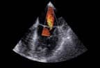
| The HD15 incorporates techology that improves workflow. |
The HD15 ultrasound system is designed to bring advanced imaging and throughput capabilities to smaller groups, including private-practice offices, clinics, small hospitals, and emerging markets. Both imaging quality and productivity are enhanced by the advanced, feature-rich technology available on the new device.
The next-generation platform is designed to overcome common challenges in interpreting images, improving accuracy and workflow. The system architecture features both the company’s proven platforms and newer technologies, including high-resolution A/D technology, 4X parallel processing, QLAB quantification software, and PureWave crystal technology.
Imaging Innovations
Philips’ PureWave technology represents a leap in ultrasound technology, featuring crystals 85% more efficient than conventional PZT material.
The extremely broad frequency range permits a single transducer to image a wide array of patients.
Company software has been equally innovative: XRES Adaptive Image Processing, SonoCT Real-Time Compound Imaging, Microfine EX focusing, and iSCAN are all available on the new system.
The company’s XRES (for extreme resolution technology) Adaptive Image Processing enhances images using proprietary algorithms designed to reduce artifacts and sharpen edges. The technology uses 350 million calculations per frame of data and is constantly adapting each pixel of the frame for each frame in time, which nearly eliminates noise.
SonoCT Real-Time Compound Imaging also improves image accuracy, reducing artifacts further. The technology creates a single compounded image at real-time frame rates by combining microangulated, coplanar, tomographic images acquired using transmit beam-steering techniques. Clinicians acquire up to nine times more information than is captured with conventional methods, according to Philips.
New company technology also improves definition. Microfine EX focusing uses new dynamic receive lens tuning with five times more focal points than older generations to improve tissue uniformity and thereby sharpen edges.
The Patient Has Left the Building
The HD15 also incorporates technology that improves workflow. Tissue Specific Imaging optimizes the transducer for the exam type automatically. Scan intelligent optimization technology, designed for two-dimensional and Doppler exams, will automatically sample digital data and adjust gain, TGC, and compression on 2D images and establish automatic scale and baseline adjustments for Doppler exams.
Active native data permits users to manipulate the exam parameters and image settings after the exam is over and the patient has left. Live compare displays previous exams against active ones so that physicians may more easily review images side by side, a feature that is particularly beneficial for interventional procedures and fetal evaluation.
The PureWave transducer technology and features, such as contrast-enhanced ultrasound, enable the device to be used to perform real-time guidance and minimally invasive procedures. Its applications also include abdominal, breast, cardiac, musculoskeletal, prostate, small parts and superficial, transesophageal, critical care, emergency medicine, ob/gyn, pediatric, intraoperative vascular, transcranial Doppler, regional anesthesia, and stress echo.
“Physicians are increasingly turning to ultrasound to help support the growing demand for diagnostic exams and treatment guidance. The HD15 brings capabilities that assure user simplicity and productivity to more clinicians, in a variety of clinical settings worldwide,” said Anne LeGrand, senior vice president of ultrasound for Philips Healthcare.
The ultrasound solution can be used as the primary or as a secondary solution. It is configurable for use as a dedicated unit or multispecialty imaging instrument, and its small size makes it an ideal bedside solution. A wide variety of configurable patient reports and exam storage options, including DVD-CD-R/RW, USB drive, and full DICOM capabilities, provide efficient patient data management as well as the opportunity for easy colleague and/or specialist consulting.
The device is intended to improve the efficiency and accuracy of ultrasound exams in all settings, particularly for more technically challenging patients and pathologies—even after patients have left the building.
—Renee DiIulio

