 |
· Putting Ultrasound Through the Paces
· Ultrasound Imaging for Electrophysiology
Putting Ultrasound Through the Paces
Teams of physicians will use the Olympic Games as a testing ground for advanced ultrasound, by examining top US athletes in studies on cardiac health and injury prevention. The project, which started as athletes trained for Beijing and will continue after the Games begin, is a follow-up to similar studies conducted at the 2006 Winter Olympics in Turin, Italy.
 |
Advancements in ultrasound technology are making the project possible, because until even just 3 years ago ultrasound technology lacked the image clarity needed for such studies, said Michael Reed, MD, US Olympic Committee medical director, performance services division. “I have a hard time describing how exciting it is to me,” Reed said. “We’re just able to look at things that we weren’t able to see before.”
With the latest round of studies, one team of physicians will seek to confirm results from Turin that showed the hearts of top athletes go through anatomic and physiological adaptations through training, and function better as a result. Another team of physicians will focus on improving the musculoskeletal health of athletes, with the aim of using close monitoring to prevent injuries.
The researchers will rely on ultrasound technology from General Electric. Physicians will take the ultrasound machines to the sidelines of Olympic competitions, to the Olympic Village, and to training centers, Reed said.
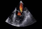
In a clinical study of weight lifters and rowers, a team led by Melissa Wood, MD, of the Massachusetts General Hospital Heart Center in Boston will use GE’s Vivid i ultrasound system to perform echocardiograms and examine athletes’ hearts before and after competition.
Wood conducted similar studies on short track speedskaters at the 2006 Winter Olympics. The speedskaters had an enlargement of the cavities of the heart and better heart function, which researchers concluded were the result of adaptations to a vigorous training regimen. Wood’s team hopes to confirm those results with athletes training for the Beijing Olympics. Physicians say the study has implications for nonathletes also.
“We have all the cardiac histories that are going in this country, and the lack of exercise and increases in body weight are leading to heart problems,” Reed said. “And you take this study and we can see that the heart can be influenced by exercise quickly and long-term, and will provide a longer, better life to people though exercise.”
—Alex Dobuzinskis
Ultrasound Imaging for Electrophysiology
Although electrophysiology is a relatively young specialty, today’s aging population and the millions who suffer from atrial fibrillation arrhythmia are drawing more attention to the need for improved therapies. More recently, clinicians have discovered that patients, in addition to feeling a whole range of systems from fatigue and dizziness, have an increased chance of having stroke.
 |
While effective, ablation—which eliminates the group of tissue that is firing inappropriately—is complex, lengthy, and therefore used infrequently. Its alternative is a lifetime of taking antiarrhythmic medication and blood thinners.
Answering the call to make atrial fibrillation a treated disease, GE Healthcare and Biosense Webster, Diamond Bar, Calif, have announced a joint agreement to develop real-time ultrasound imaging for use in electrophysiology procedures.
The collaboration is a “natural extension” of GE’s long-standing technology partnership with Biosense Webster, explained Mark Langer, general manager of GE Healthcare’s Interventional Cardiovascular Ultrasound division. About 6 years ago, the companies discovered there would be a real benefit in integrating GE’s CardioLab IT Recording System with Biosense Webster’s CARTO XP Navigation and Ablation System. Two years later, the pair introduced the bidirectional interface, which allowed clinicians to automatically share recording and mapping information, synchronize data, and deliver a single, comprehensive EP case report with events, vitals, waveforms, ablation parameters, and CARTO 3D electroanatomical maps. In what Langer described as a “great result, and a great step forward” for the companies, more than 300 health institutions have enjoyed the solution and its ability to eliminate the disparity that would traditionally come with two disconnected systems.
—Elaine Sanchez


