|
· On the Road with Digital Mammo
· Partners in Progress: Fujifilm Scores Deal with Premier for DR
· Teaming Up on X-ray Tubes
· Product Alert: Varian Launches New Multileaf Collimator for IMRS
On the Road with Digital Mammo
In today’s world, women are becoming increasingly educated on the benefits of digital mammography, so much so that female patients are requesting it themselves.
This is a trend that DMS Imaging, a member of DMS Health Group, headquartered in Fargo, ND, heard from some of its clientele. When the company sent out a letter asking if any others had an interest in the advanced technology, it received an explosive response.
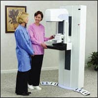 |
| DMS Imaging recently added full-field digital mammography to its list of mobile services. |
To satisfy this demand, DMS Imaging decided to take its expertise on the road. The company has recently added full-field digital mammography to its list of mobile services.
Corey Nesemeir, account executive for DMS, said one of the driving forces for digital mammography replacing traditional analog routes is that facilities are looking to replace equipment and optimize space. It allows radiologists to view the x-ray image more closely through zoom features, it presents a better way of viewing calcifications in the breast masses, and it reduces the amount of time it takes to review the images by more than one half. “The efficiency is drastically improved,” Nesemeir said.
Nathan Roesler, DMS Imaging regional vice president, also described how through shared service pooling, the company is able to bring direct-capture digital mammography to the rural community, where even larger facilities cannot afford it within their walls. The concept of the shared model incorporates economic resources of many facilities, in order to provide cutting-edge medical services to the people in their communities, Roesler continued. DMS currently has just under 20 rural health care clients in the shared model.
“Part of the mission of DMS Imaging is that we believe all people, no matter where you live, deserve cutting-edge health care services,” Roesler said, adding that the company’s objective is to export its services even further on a national level. “Our goal is to deliver this product and service to any customer base out there, from coast to coast, who’s interested in contracting the service. It appears that’s a goal that we can attain.”
DMS Imaging currently operates one analog unit, with two digital routes slated for North Dakota. It has been in operation for more than 25 years, originating in Minnesota and expanding to a national scale. The company provides mobile services in a variety of modalities, including MRI, CT, ultrasound, and nuclear medicine.
—Elaine Sanchez
Partners in Progress: Fujifilm Scores Deal with Premier for DR
Joins five others in newly awarded contracts
Premier Purchasing Partners LP, the group purchasing division of Premier Inc, recently selected Fujifilm as a contracted supplier for direct radiography.
Listed in the contract is Fujifilm’s complete product line of DR table and upright systems, including the company’s Velocity SpeedSuite.
 |
According to Fujifilm, the company’s direct radiography offerings are similar, but unique.
DR detectors provide the highest resolution and fastest image availability of any DR system on the market, along with the lowest cost of ownership over the life of the system.
“Premier is a valued partner, and we’re pleased to be able to provide their members with leading products that will ultimately enhance their patient care,” said Phil Buffington, Fujifilm’s executive director of national accounts. “The quality, reliability, and productivity of Fujifilm’s innovative DR systems are continually evolving, resulting in not only enhanced care, but even greater operational efficiencies as well.”
Introduced during last year’s RSNA meeting, the Velocity SpeedSuite includes the Velocity-Ufp, a compact reader for chest and other upright exams that provides throughput of up to 240 images per hour, in addition to a Velocity-Tfp table system, designed to accommodate increased patient weights up to 506 pounds.
Premier will also receive the SpeedSuite Unity, a fully motorized, flexible U-arm system that is ideal for performing a full range of imaging examinations both in the hospital setting as well as outside the hospital in imaging centers and clinics.
While he refrained from commenting on any one contract, Premier Vice President for Sourcing Mike Georgulis said bidding periods generally take about 9 months, depending upon the nature of specific categories. This period calls for product planning, product cross referencing, and input from its member clinical committees through face-to-face and conference-call discussions.
The ultimate decision is based on clinical efficacy, cost of products, manufacturing capacity, sales force support, distribution methods, and other company attributes, Georgulis continued. When it comes to cost, that component is uniquely defined for each category.
This derived information may facilitate the monitoring of stages of disease in individual patients.
Among its various features, NeuroQuant first minimizes scanner image-acquisition artifacts that formerly hindered automated MRI analysis. After the initial screening, intelligent image-analysis and feature-extraction processes identify anatomical structures in the data that are known to atrophy in particular diseases. Once numerical information about the sizes of the structures is extracted, the analysis is presented to the referring physician in the form of an easy-to-read report. Ultimately, the document’s contents can be compared to age-appropriate normative data.
University of California, San Diego, neurologist James Brewer, who studies the effects of Alzheimer’s disease on the brain, said postprocessing MRI data with NeuroQuant provides an efficient means of evaluating treatment options while considering the measurable extent and progression of patient brain atrophy.
“There is a great deal of scientific evidence that brain structures, such as the hippocampus, begin to atrophy in the early stages of dementia,” Brewer said. “Reliably characterizing such changes based on MRI data requires quantitative techniques, but previous methods for deriving numerical information from MRI scans have been too slow and cumbersome for use outside of research studies.”
The software product has received 510(k) clearance from the FDA for marketing as a medical device “intended for automatic labeling, visualization, and volumetric quantification of segmentable brain structures from a set of MR images.”
The MRI scans routed from a scanner or PACS are received as input, and NeuroQuant can automatically return age-related atrophy reports and numerical and color-blended anatomical volumes annotated with graphical overlays to most DICOM-compliant PACS viewers or third-party workstations. Additionally, the data-analysis tool can be configured to supply automated MRI quantification solutions to independent health care providers, imaging centers, and large firms conducting multisite clinical trials.
CorTechs President Michael E. Smith praised the team of engineers and researchers who created the data-analysis software, noting the resource’s self-sufficient components.
“CorTechs’ outstanding engineering team and affiliated researchers have developed a powerful pipeline of sophisticated analysis processes that can quantify MRIs without human intervention,” Smith said.
This derived information may facilitate the monitoring of stages of disease in individual patients.
—E. Sanchez
Teaming Up on X-ray Tubes
Siemens Medical Solutions USA Inc, Malvern, Pa, and Xintek Inc, Research Triangle Park, NC, recently announced the formation of a joint venture company called XinRay Systems. The purpose of the company is to develop a new multipixel x-ray source technology for a range of diagnostic imaging applications.
“X-ray is a mature technology,” noted Xintek chairman Otto Zhou. “It has been around for a long time, over 100 years now. With today’s x-ray tubes you have a hot filament you run at 1,000 degrees or so to generate electrons. And all x-ray tubes have what we call a single pixel design.”
XinRay’s x-ray tubes will work differently, explains Zhou. “The technology we’re working on is nanotechnology based,” he said. “We generate electrons at room temperature, and those are excited with an electrical field. We can fabricate them much more easily, and that allows us to create a multipixel x-ray source with information coming in at different points.”
 |
This technology could be very powerful because it will have the ability to generate images from different angles without device rotation. In the short term, says Zhou, XinRay hopes to develop more compact and efficient equipment for use in security situations, including luggage screening at airports. In the long term, those same advantages will be brought to bear on a new generation of imaging equipment, beginning with digital mammography devices.
“With nonmedical applications and low-current medical applications like mammography, we expect to have products hitting the market in the next few years,” Zhou said. “In other areas, there are still more challenges to overcome. With some medical imaging equipment, the x-ray power requirement is very high, so we need a substantial amount of development on our part to overcome the technical challenges and make sure the source gives the performance required. But that’s not the case for low-power x-ray applications.”
XinRay combines Siemens and Xintek research and development under one roof in Research Triangle Park; its staff is drawn from both Xintek and Siemens facilities in Germany and China.
“Siemens has been producing x-ray tubes for a long time, so there is a solid background and good knowledge of the technology,” noted Moritz Beckmann, who was originally drawn from Siemens in Germany and is now COO of the up-and-running joint venture. “Siemens is very experienced in quality management as well as manufacturing control and logistics, and we bring in experience in registration and certification of new products.”
And Xintek brings its specialization in nanotechnology to the table. “Xintek is a university start-up that spent several years developing this technology,” Zhou said. “Now we have experience fabricating the high-performance field emission cathode source.”
The company’s tubes for digital mammography systems, utilizing the new source technology, could be on the market in just a few years, Zhou says. “We have a system where we can generate multiple views without mechanical motion,” he said. “It will enable faster scanning time and reduce the level of compression-related discomfort for the patient during imaging.”
Improved resolution would be another benefit of the technology, Zhou noted. “You have spatially distributed x-ray radiation that can be turned on in a sequence,” he said. “That’s the bottom line of this new technology.”
—Cat Vasko
 |
| Positioned for a frameless stereotactic radiosurgery treatment beneath the multileaf collimator, which shapes the radiation beams during treatment. |
Product Alert: Varian Launches New Multileaf Collimator for IMRS
While statistics on intensity-modulated radiosurgery is currently difficult to come by, numerous studies refer to IMRS’ emergence as one of the latest advances in radiology. For example, a March 2007 article in Neurosurgery Quarterly discusses spinal stereotactic radiosurgery as a promising treatment option for metastatic epidural spinal cord compression.
Recognizing the necessity of precision in what is becoming widely known as an effective approach to treatment of cancer and other indications, Varian Medical Systems Inc, Palo Alto, Calif, has created an ultra-fine beam-shaping device for radiosurgery called the HD120 MLC multileaf collimator. The new medical device received FDA 510(K) clearance in September.
Included in the Novalis Tx radiosurgery system that resulted out of Varian’s partnership with BrainLAB, Westchester, Ill, the collimator is one component of a noninvasive alternative to conventional surgery. Specifically, it will be used to treat a variety of conditions, including benign and malignant tumors, metastatic cancer, arteriovenous malformations, trigeminal neuralgia, and acoustic schwannoma.
“Successful irradiation of these types of abnormalities depends on doctors’ ability to deliver a high dose of radiation with the precision of a surgical strike, sparing the surrounding tissues,” said Calvin Huntzinger, marketing and engineering manager for Varian’s Surgical Sciences product line. “Varian’s new HD120 MLC multileaf collimator doubles the resolution of our best MLC, so it is ideal for carrying out delicate intensity-modulated radiosurgeries near critical structures like the optic chiasm, brain stem, or spinal cord.”
The multileaf collimator uses computer-controlled mechanical leaves to continually shape the treatment beam while radiation is delivered from different angles around the patient. Compared to Varian’s previous line of multileaf collimators, the company’s latest model sports central leaves whose width has been sliced in half, at 2.5 mm instead of 5. This reduction increases beam-shaping precision by 100 percent, according to Varian.
Also, the new product features improved dose delivery capabilities, such as a steeper dose fall-off gradient. Because the dosage amount decreases rapidly outside the targeted area, healthy tissues are better protected, Varian officials say.
“The multileaf collimator has revolutionized radiotherapy, and it is now poised to do the same with radiosurgery,” Huntzinger said. “We are committed to extending this life-saving technology to help more patients in a broader range of cases.”
—E. Sanchez