As part of the “Thematic session 10: New technology for urology” plenary session at the European Association of Urology (EAU) Virtual Annual Meeting, Wietske van der Zwaag, PhD, discussed new imaging approaches in urology, focusing on the 7T MRI.
All magnetic resonance imaging (MRI) systems contain a very large magnet. In the clinical setting thus far, these are typically 1.5 or 3 Tesla in strength. Magnets of 7 Tesla or higher are deemed ultra-high field. To date, there are relatively few of these, with approximately 80 to 100 worldwide.
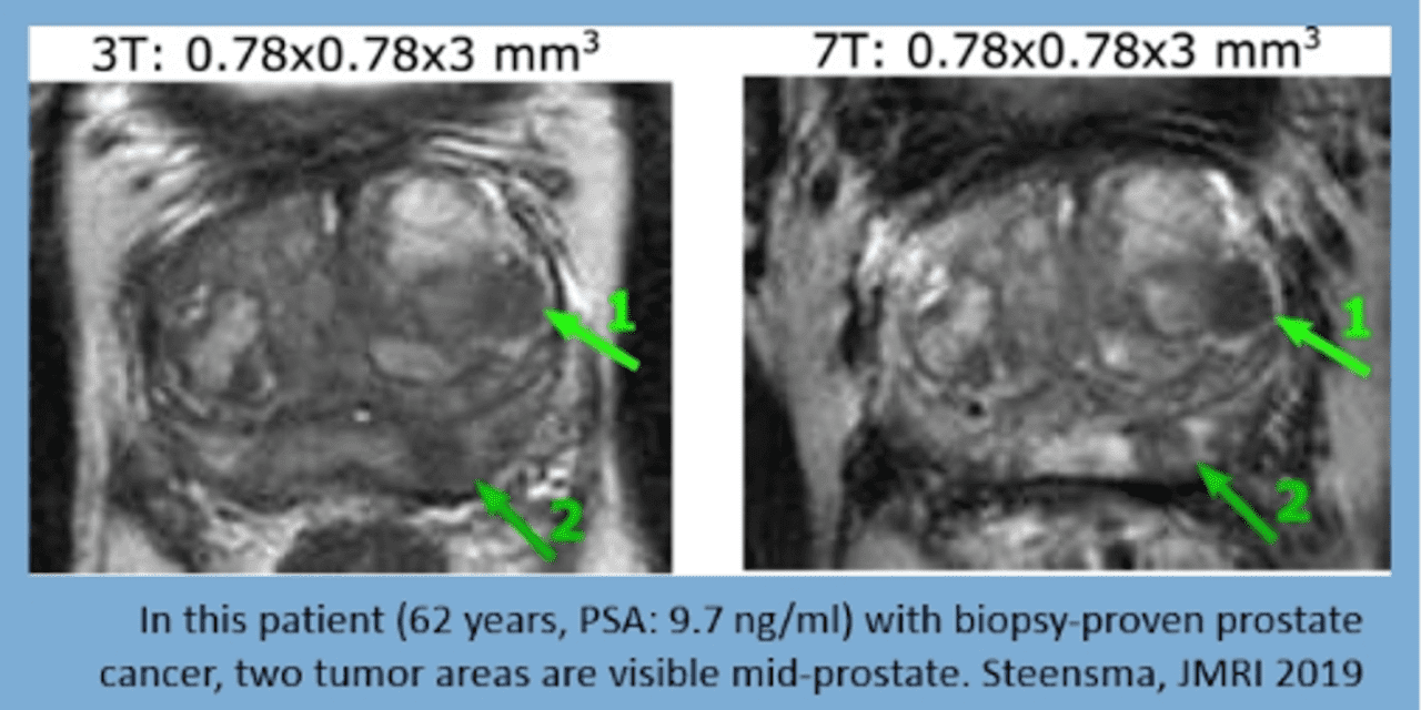
The most important benefit of a transition from standard to the ultra-high field is an increase in the signal-to-noise ratio, which allows for more detailed assessment, as highlighted in the accompanying figure.
Ultra-high-field MRI also has the ability to maximize benefit in functional MRI due to a linear increase in signal intensity with increasing field strength. This higher signal can be utilized to improve spatial resolution, allowing for the subdivision of anatomic regions.
While many of these uses focus on neuro-imaging, this has potential benefits in urology. van der Zwaag highlighted the assessment of pelvic muscle contraction and tongue movement, two midline, voluntary motor tasks. They demonstrated two pelvic muscle clusters. Such an approach is also feasible in terms of a sensory input using tactile feedback, as demonstrated with an assessment of the penile shaft and left and right feet. Again, two shaft clusters were identified.
Using individual MRI imaging, investigators can determine MRI-based biomarkers on the basis of the extent of what is active, the distance between clusters, and timecourse properties.
7T MRI can provide high-resolution images with the ability to map brain activation at an individual level.
Presented by: Wietske van der Zwaag, PhD, Spinoza Centre for Neuroimaging, Amsterdam, Netherlands
Written by: Christopher J.D. Wallis, Urologic Oncology Fellow, Vanderbilt University Medical Center, @WallisCJD on Twitter, at the Virtual 2020 EAU Annual Meeting #EAU20, July 17-19, 2020.
Featured Image: In this patient with biopsy-proven prostate cancer, two tumor areas are visible mid-prostate. You can see 3T MRI and 7T MRI images side by side. (Credit: Steensma, JMRI 2019)