Summary: University of California, San Francisco researchers developed a machine learning algorithm that enhances 3T MRI images to simulate 7T resolution, improving the detection of brain abnormalities like those in traumatic brain injury and multiple sclerosis, though further clinical validation is needed.
Key Takeaways
- Need for Further Validation: While the initial results are promising, the researchers emphasize the importance of clinical assessments, expert evaluations, and further testing before the model can be integrated into routine clinical practice.
- AI-Enhanced MRI Imaging: Researchers at the University of California, San Francisco developed a machine learning algorithm that generates 7T-like images from standard 3T MRIs, improving the clarity and detection of brain abnormalities.
- Clinical Application Potential: The model enhances visualization of conditions like traumatic brain injury and multiple sclerosis by revealing clearer contours of lesions and microbleeds that are harder to detect with conventional imaging.
—————————————————————————————————————————————————————————————————
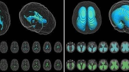
University of California, San Francisco (UCSF) researchers have created a machine learning algorithm to generate synthetic 7T MRI images from standard 3T scans. Their work, presented at the 27th International Conference on Medical Image Computing and Computer-Assisted Intervention (MICCAI), aims to improve the detection of brain abnormalities, including those found in traumatic brain injury (TBI) and multiple sclerosis (MS).
“Our model synthesizes higher-quality MRIs from lower-field systems, improving the clarity of brain abnormalities,” says Reza Abbasi-Asl, PhD, assistant Professor of Neurology at UCSF. “We show how AI can enhance the visibility of subtle changes in the brain that are harder to detect with conventional imaging.”
Transforming 3T MRIs into 7T-like Images
While 1.5T and 3T MRIs are common in clinical settings, 7T MRIs offer superior resolution. However, fewer than 100 7T machines are available globally, limiting access. UCSF’s model uses 3T MRIs to generate 7T-like images, providing a more detailed view of brain tissue. The study focused on mild TBI cases, with the AI-enhanced images revealing clearer contours of subcortical microbleeds and better separation between white matter lesions.
“These synthesized images offer improved visualization of pathological tissue,” Abbasi-Asl says. “We found the generated 7T images gave better insights into white matter lesions and microbleeds, helping differentiate between adjacent lesions and highlighting key features.”
The researchers stress the need for further validation before these models can be integrated into clinical practice. Future studies will include clinical assessments, ratings from medical professionals, and further analysis to evaluate the accuracy and reliability of the AI-generated images.