The 2023 Radiological Society of North America (RSNA) Alexander R. Margulis Award for Scientific Excellence award will be presented to the authors of the Radiology article “Pancreatic Cancer Detection on CT Scans with Deep Learning” during the RSNA 2023 annual meeting in Chicago from November 26-30.
Named for Alexander R. Margulis, M.D., an investigator and visionary in the science of radiology, this annual award recognizes the best original scientific article published in RSNA’s flagship journal, Radiology.
“This year’s Margulis Award recognizes impactful results likely to affect millions of patients throughout the world,” says Radiology Editor Linda Moy, MD. “The study demonstrates how a deep learning-based tool can result in accurate detection of pancreatic cancer on CT scans, especially for tumors smaller than two centimeters. Early detection of pancreatic cancer allows for prompt intervention that greatly increases the chances of survival.”
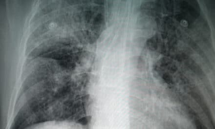
Pancreatic cancer patients face a poor prognosis, with a five-year survival rate of only 12%, according to the American Cancer Society. Early detection is the best way to improve the odds. Prognosis worsens significantly once the tumor grows beyond two centimeters and spreads outside the pancreas.
CT, the most widely used and most sensitive exam for pancreatic cancer detection, misses about 40% of tumors smaller than two centimeters. A tool to boost pancreatic cancer detection is urgently needed.
For the study, Po-Ting Chen, MD, co-lead author Tinghui Wu, MS, and colleagues from National Taiwan University in Taipei, Taiwan, developed an artificial intelligence (AI) deep learning tool and trained it by comparing hundreds of contrast-enhanced CT exams from patients with and without pancreatic cancer.
The AI tool in the study achieved 90% sensitivity and 93% specificity in a test set of 1,473 real world CT exams. Sensitivity was comparable with that of radiologists regardless of tumor size and stage. Sensitivity for detecting pancreatic cancers less than two centimeters was 75%.
“In terms of early detection and diagnosis, our workflow plays a pivotal role in identifying pancreatic cancer at earlier and more treatable stages,” Chen says. “By aiding radiologists and clinicians in recognizing suspicious lesions on CT scans, it facilitates swift and accurate diagnosis, which is crucial for improving patient outcomes. Furthermore, this workflow offers a valuable advantage by providing a reliable second opinion, enhancing diagnostic confidence among medical professionals, and ultimately benefiting patient care.”
Importantly, the method uses automated pre-processing segmentation, or identification and outlining of the pancreas on whole-body CT scans. Automation of this process represents an important advance in AI evaluation of pancreas imaging, as the pancreas borders multiple organs and structures and varies widely in shape and size.
“This approach not only streamlines the process, saving valuable time for physicians that would otherwise be spent manually delineating the region of interest, but it also ensures that the classification model is directed toward the critical area, eliminating extraneous information,” Chen says.
Computer-aided segmentation also enables quantitative analysis including measuring the size, shape and volume of the pancreas and any detected lesions, aiding in treatment planning and disease monitoring.
“We are truly honored and genuinely surprised to have received this award,” Chen says. “It’s a recognition of the hard work and dedication that our team has put into our research. We want to express our deep gratitude to the award committee for this incredible acknowledgment.”