Experts highlight benefits of 3D SVR MRI in visualizing fetal optic pathway
A Scientific Online Poster that was presented during the 2023 ARRS Annual Meeting on the island of Oahu, Hawaii, reportedly explained that the technique of three-dimensional (3D) slice-to-volume (SVR) MRI allows for precise delineation and measurement of the fetal optic pathway (FOP).
During the presentation, experts noted the limited fetal presentation and low reproducibility of ultrasound-based techniques, as well as conventional MRI’s inconsistencies in FOP visualization due to low resolution.
“Our preliminary results nevertheless demonstrate the promises and utility of this technique,” explained Eric Juang, MS, of Creighton University School of Medicine’s Phoenix Regional Campus and Phoenix Children’s Hospital in Arizona.
Juang and his team explained that in their study, all fetal MRI examinations performed at Phoenix Children’s Hospital between January 1, 2020 and August 1, 2022 were reviewed to find those with sufficient quality to reconstruct a 3D SVR image. They reported that a medical student reader first examined the unprocessed fetal brain MRI—either from balanced turbo-field-echo or T2-weighted single-shot fast spin echo (T2 SSFSE) sequences—attempting FOP measurements where feasible. They further explained that 3D SVR reconstructions of fetal brain images were performed using a minimum of six T2 SSFSE imaging sequences. With that same reader next examining the reconstructed imaging and recording FOP measurements, two pediatric neuroradiologists read all FOP measurements, and to estimate the relationship between FOP measurements of normal fetuses and gestational age, nomograms were generated accordingly.
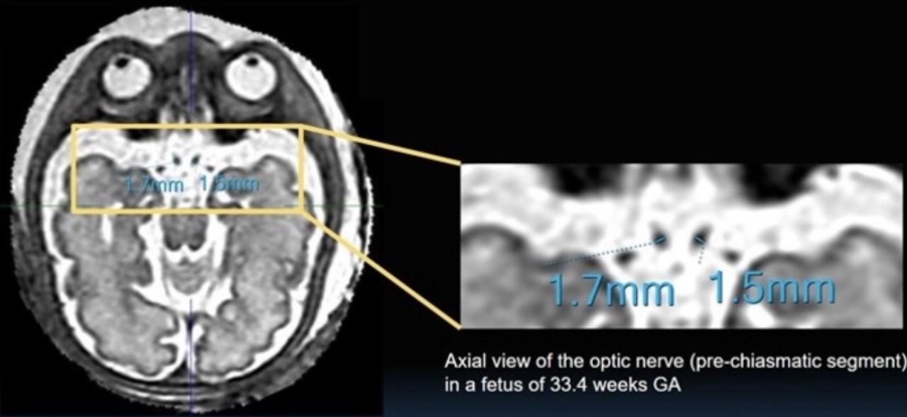
The team went on to explain that out of 70 fetal MRI scans selected for this ARRS Annual Meeting Summa Cum Laude Online Poster, FOP was visualized in 9 cases in unprocessed fetal MRIs, compared to 55 cases in 3D SVR images. According to the researchers, among the 55 3D SVR cases, pre-chiasmatic optic nerve width was successfully measured bilaterally in 53 cases, optic chiasm width in all 56 cases, and bilateral optic tract width in 30 cases.
Specifically, a linear regression fit estimated the relationship between optic chiasm width (OCW) in millimeters in normal fetuses and gestational age (GA) in weeks as OCW = 0.11 × GA+2.0 (R^2 = 0.30); similarly, the relationship between pre-chiasmatic (PC) optic nerve width and GA was estimated as PC = 0.04 × GA +0.24 (R^2 = 0.34), experts reported.
“Further results are pending,” Juang and his team added—reiterating their belief that early detection of FOP defects remains critical for improving patient outcomes.