Summary: X-ray–induced acoustic CT enables 3D imaging from a single X-ray projection, reducing radiation exposure, improving efficiency, and offering a compact alternative to traditional CT systems.
Key Takeaways
- XACT Enables 3D Imaging with a Single X-Ray Projection: Unlike traditional CT, which requires hundreds of projections, XACT uses X-ray-induced acoustic signals to achieve 3D imaging efficiently.
- Reduced Radiation and Compact Design: XACT significantly lowers radiation exposure and eliminates the need for large, complex gantry systems, making it safer and more versatile.
- Potential for Medical and Industrial Applications: XACT’s innovative approach unlocks new possibilities for imaging in constrained environments, revolutionizing medical diagnostics and nondestructive testing in engineering and material science.
———————————————————————————————————————————————————
CT has been a cornerstone of imaging, providing detailed 3D insights into the human body and materials. Traditional CT, however, requires hundreds of X-ray projections from multiple angles, leading to significant radiation exposure and reliance on large, stationary systems.
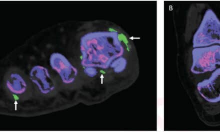
To address these limitations, researchers from UC Irvine‘s Departments of Radiological Sciences and Biomedical Engineering have developed a new technology, X-ray–Induced Acoustic CT (XACT). Published in Science Advances, XACT enables 3D imaging using just a single X-ray projection, reducing radiation exposure and offering a more efficient alternative.
A New Paradigm in Imaging
“In XACT, the generated sound waves by X-rays change the way X-ray imaging works, converting X-rays to ultrasound. X-rays typically travel in straight lines, so one projection only provides 2D information. However, X-ray-induced acoustic signals propagate in three dimensions, allowing for 3D imaging with a single projection,” says Shawn Xiang, PhD, the study’s corresponding author and an associate professor at UCI’s Departments of Radiological Sciences and Biomedical Engineering.
XACT leverages the interaction between X-rays and tissue to produce acoustic waves, which travel at a speed of 1,500 meters per second. These waves are captured by ultrasound detectors, enabling real-time, 3D imaging without the need for mechanical scanning or complex gantry systems.
“For the first time, we have proved that 3D imaging can be obtained with a single X-ray projection based on X-ray-induced acoustic detection in both phantoms and biological tissue,” says Siqi Wang, PhD, the study’s first author. Wang completed his PhD at UCI in Xiang’s lab and is now a postdoctoral research scholar at Stanford University.
“The groundbreaking finding here is that you can make 3D X-ray imaging with just a single projection, which typically needs 600 projections or more,” adds Vahid Yaghmai, MD, MS, FSAR, a radiologist at UC Irvine and chair of the UC Irvine Department of Radiological Sciences, who was not directly involved in the study.
Benefits Beyond Traditional CT
One of XACT’s most significant advantages is its efficiency and reduced radiation exposure. This makes XACT a safer and more accessible alternative, particularly for applications like routine diagnostics and breast cancer screening. Furthermore, with portable X-ray sources and ultrasound detectors, XACT systems promise compact, gantry-free designs, enabling imaging in settings previously inaccessible to traditional CT systems.
Challenges and Future Directions
While the potential of XACT is immense, current limitations include resolution constraints tied to the frequency and size of the ultrasound detectors. Future improvements, such as higher-frequency transducers and advanced reconstruction algorithms powered by deep learning, could further enhance its performance.
Redefining Imaging Across Fields
According to UC Davis experts, XACT enables 3D imaging from a single X-ray projection, revolutionizing both medical diagnostics and nondestructive testing in engineering and material science. By eliminating the need for rotational access, XACT unlocks imaging possibilities in constrained environments.
The researchers emphasize that this technology marks a significant leap forward, offering reduced radiation exposure, a compact design, and exceptional efficiency. As XACT continues to evolve, they believe it has the potential to redefine medical and industrial imaging, paving the way for high-resolution, low-dose 3D imaging to become the standard in healthcare and beyond.