One in 8 women in the United States will develop breast cancer during their lifetime, and more than 40,000 die from the disease annually, according to the Centers for Disease Control and Prevention.
Early detection and effective treatment are the keys to preventing deaths. Advances by University of Texas at Dallas researchers on multiple fronts show promise for helping detect and monitor the disease more accurately, better assessing women’s risk and attacking treatment-resistant tumors.
Hyperspectral Imaging
UT Dallas engineers are applying artificial intelligence (AI) to imaging technologies to develop advanced tools to identify cancer cells, including noninvasive methods that do not expose patients to radiation or contrast agents.
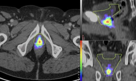
Hyperspectral imaging (HSI), for example, has the potential to help doctors identify cancerous cells with more accuracy. HSI, originally used in satellite imagery, examines cells under ultraviolet and near-infrared lights at micrometer resolution, producing spectral images as unique as fingerprints. UT Dallas researchers have combined HSI with deep learning, a machine learning technique, to compare images automatically with others in an extensive database and help doctors distinguish between normal and cancerous cells.
After publishing research in 2019 that demonstrated the technology could detect cancer cells in head and neck cancer surgery patients, Dr. Baowei Fei, professor of bioengineering, and his team recently applied the technique to breast cancer. They presented their study at the SPIE, the international society for optics and photonics, conference in February in Houston.
“If we have a large database that knows what is normal tissue and what is cancerous tissue, then we can train our system to learn the features of the spectra and predict whether a sample is cancerous or not,” says Fei, the Cecil H. and Ida Green Chair in Systems Biology Science in the Erik Jonsson School of Engineering and Computer Science. He has a joint appointment as a professor of radiology at UT Southwestern Medical Center.
The research was funded in part by the Cancer Prevention and Research Institute of Texas (CPRIT).
AI and Mammograms
Researchers in the Jonsson School’s Quality of Life Technology Labs have built an AI system that can scan volumes of mammograms and accurately identify cancer as well as determine breast density. They presented research on using AI to predict breast density in July at the 42nd annual International Conference of the IEEE Engineering in Medicine and Biology Society and published a study about their approach to scan mammograms in the April 2019 issue of Computers in Biology and Medicine.
“Breast cancer is the second-leading cause of cancer death for women, and there is a shortage of mammography professionals around the world, which threatens the timely care for those affected. Our AI system can help doctors provide timely care more cost effectively,” said Dr. Lakshman Tamil, a professor of electrical and computer engineering and the lab’s director.
Thermal Imaging
Dr. Fatemeh Hassanipour, associate professor of mechanical engineering, and fellow researchers recently built a proof-of-concept computer model of the thermal properties of breast cancer, a critical step toward making digital infrared thermal imaging more useful for monitoring breast cancer.
Her research, conducted with UT Southwestern radiologists, was published June 22 in Nature Research’s Scientific Reports.
Hassanipour’s work is supported by a National Science Foundation Faculty Early Career Development Program (CAREER) award, which she received in 2015 to study the biomechanics of breastfeeding.
“Infrared imaging could potentially provide useful information in a diagnostic setting to radiologists,” Hassanipour said. “We want it to be used like a second device for monitoring tumors.”
Online Risk Assessment
An online tool developed by statisticians in the School of Natural Sciences and Mathematics helps breast cancer patients assess their future risk of getting contralateral breast cancer – the development of cancer in the other, healthy breast. The tool can help doctors determine the best course of action, such as whether to remove the healthy breast.
“This project really shows the important role mathematics and statistics play in areas vital to our everyday lives,” said Dr. Swati Biswas, professor of mathematical sciences. The research was funded by the National Cancer Institute.
Molecule for Treatment-Resistant Cancer
Dr. Jung-Mo Ahn, associate professor of chemistry, designed a small molecule, ERX-11, that could help breast cancer patients for whom current treatments no longer work.
In a paper published in 2017 in the online journal eLife, Ahn and his colleagues describe their approach to designing the molecule as well as experiments that show its effectiveness at stopping the progression of treatment-resistant breast cancer cells in isolation and in an animal model. Funding sources for the research included CPRIT, the National Institutes of Health and The Welch Foundation.
“Eventually we might be able to develop molecules similar to ERX-11 to treat other types of cancer as well,” Ahn says.