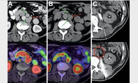
PSMA PET-CT offers a more accurate way to stage men with high-risk localized prostate cancer than conventional imaging, according to a recent study reported in Renal and Urology News.
“PSMA PET-CT is a suitable replacement for conventional imaging, providing superior accuracy, to the combined findings of CT and bone scanning,” Michael S. Hofman, MBBS, of the Peter MacCallum Cancer Centre in Victoria, Australia, and colleagues concluded in a paper published in The Lancet.
In a phase 3 trial, Dr Hofman and his collaborators studied 300 men with high-risk localized prostate cancer being considered for radical prostatectomy or radiotherapy with curative intent. Investigators randomly assigned the men to undergo conventional imaging with CT and bone scans (152 patients) or gallium-68 PSMA-11 PET-CT (148 patients). The study had a crossover design whereby men who had no more than 2 unequivocal distant metastases on first-line imaging underwent second-line imaging with the other imaging modality.
PSMA PET-CT demonstrated 27% greater accuracy than conventional imaging (92% vs 65%) in detecting pelvic nodal or distant metastases, the investigators reported. Compared with conventional imaging, PSMA PET-CT had greater sensitivity (85% vs 38%) and specificity (98% vs 91%).
Read more from Renal and Urology News and find the study in Lancet.


