Researchers from Purdue University have developed a new imaging technology that may shed more light on tumors in the body and help understand how certain diseases affect brain activity.
“We are using light to extract new information from tissue to inform doctors and assist them in designing and carrying out surgeries to remove tumors,” says Brian Bentz, PhD, who worked on the technology with Kevin Webb, PhD, a professor of electrical and computer engineering at Purdue. “It is a localization method where our technology helps the surgeon pinpoint precise information about the depth and location of tumors. Such information is not easily accessible with current technologies.”
The Purdue technology uses contrast in the absorption of light and fluorescent agents that are introduced into the body to find tumors and/or blood vessels within the tissue. The same technology can be used to study neuron activation in the brain, which can help doctors detect diseases such as Parkinson’s.
Bentz said the Purdue technology overcomes one of the major challenges with fluorescence imaging—the light becomes highly scattered and that limits the information that a surgeon receives.
“Our technology aims to provide more detailed information about tumors for surgeons and neuron activity in the brain, both of which can improve outcomes for patients,” Bentz said.
Read more from Purdue University and find the study at IEEE Transactions on Medical Imaging.
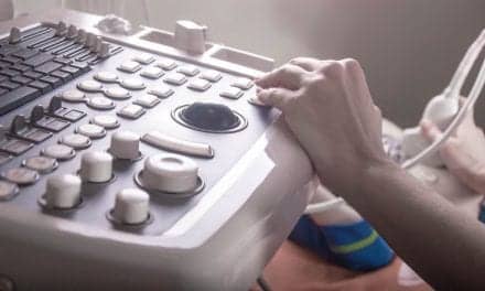
Featured image: A new tool for medical professionals may help shed more light on tumors in the body and how the brain operates. Purdue University researchers created technology that uses optical imaging to better help surgeons map out tumors in the body and help them understand how certain diseases affect activity in the brain. Credit: Purdue University/Brian Bentz.