The novel radiopharmaceutical 18F-PSMA-1007 is effective for detecting malignant prostate cancer lesions and readily available, according to research published in the April issue of The Journal of Nuclear Medicine. With this new agent and 68Ga-PSMA-11, which is already widely used, nuclear medicine departments will now have two options for staging of prostate cancer, potentially increasing availability for patients worldwide.
68Ga-PSMA is a PSMA-labeled tracer commonly used in clinical practice around the world for evaluating extent of disease in prostate cancer patients. However, 68Ga has a half-life of only 68 minutes (vs 110 min for 18F) and emits a higher-energy positron than 18F. “The newly introduced 18F-PSMA-1007 has several advantages over the established 68Ga-PSMA,” says Einat Even-Sapir, MD, PhD, director of The Institute for Nuclear Medicine at Tel Aviv Sourasky Medical Center in Israel. “These include a longer half-life, favorable pharmacokinetics, central mass production and potentially better spatial resolution.”
The prospective study compared the diagnostic accuracy of 18F-PSMA-1007 with 68Ga-PSMA-11 PET/ CT in the same patients presenting with newly diagnosed intermediate- or high-risk prostate cancer. 18F-PSMA-1007 and 68Ga-PSMA-11 PET/CT were performed within 15 days of each other in 16 patients who were scheduled to undergo a radical prostatectomy. Findings from the two PET tracers were compared with histopathologic findings obtained from radical prostatectomy specimens, considered the gold standard.
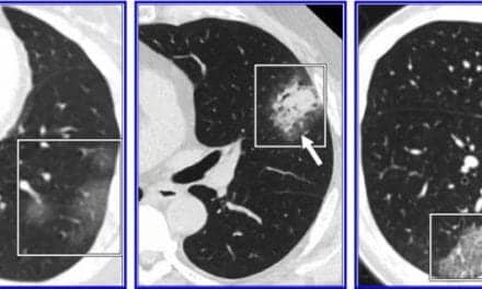
Radiolabeled PSMA-avid lesions in the prostate were identified in all 16 patients with almost perfect agreement between the two tracers regarding tumor location. Additionally, in four patients, a second positive focus, though less intense, was detected only by 18F-PSMA-1007. Three of these secondary foci were confirmed as areas of prostate cancer, while the fourth was shown on pathological examination to represent chronic prostatitis.
“In view of the near-equal performance of the two tracers, this preliminary study suggests the routine use of 18F-PSMA-1007 in lieu of 68Ga-PSMA-11 for staging prostate cancer patients; clinicians can use either radiotracer based on availability,” says Jonathan Kuten, MD, MHA, nuclear medicine specialist at Tel Aviv Sourasky Medical Center in Israel. “It also adds to the collective growing database of evidence supporting the use of PSMA agents for staging intermediate and high-risk patients. We encourage researchers, in the future, to corroborate this study’s results in larger cohorts.”
Find the study in The Journal of Nuclear Medicine.
Featured image: Maximum-intensity projections, transaxial fusion, and PET images of 18F-PSMA1007 (A–C) and 68Ga-PSMA-11 (D–F) PET/CT scans of 67-y-old patient with GS 8 and PSA 4.9 ng/mL. Marked uptake is seen in urinary bladder and left ureter (arrow) on maximum-intensity projection image of 68Ga-PSMA-11 (D), as opposed to nearly negligible 18F-PSMA-1007 urinary excretion (A). Dominant lesion in left prostatic lobe is evident on both scans (arrowheads). However, second lesion is seen in right lobe only on 18F-PSMA-1007 scan (arrow in C), later verified on pathology as true malignant lesion. Courtesy, SNMMI.