Researchers have developed a new intravascular imaging technique that could one day be used to detect coronary plaques that are likely to lead to a heart attack. Heart attacks are often triggered when an unstable plaque ruptures and then blocks a major artery that carries blood and oxygen to the heart.
“If unstable coronary plaques could be detected before they rupture, pharmacological or other treatments could be initiated early to prevent heart attacks and save lives,” says research team leader Seemantini Nadkarni, PhD, from the Wellman Center for Photomedicine at Massachusetts General Hospital in Boston. “Our new imaging technique represents a major step toward achieving this.”
In the journal Biomedical Optics Express, the researchers report a preclinical demonstration of their new intravascular laser speckle imaging (ILSI) technique in a living animal model. They show, for the first time, that ILSI can identify the distinct mechanical features of plaques that are most likely to rupture under physiological conditions of cardiac motion, blood flow, and breathing.
“Reducing mortality from heart attacks in the general population requires a comprehensive screening strategy to identify at-risk patients and detect high-risk vulnerable plaques while they can be treated,” says Nadkarni. “By providing the unique capability to measure mechanical stability—a critical metric in detecting unstable plaques—ILSI is poised to provide a new approach for coronary assessment.”
Although intravascular technologies have been developed to evaluate microstructural features of unstable plaques, recent studies have shown that mechanical features, in addition to microstructural and compositional features, influence plaque rupture.
“Measurement of the plaque mechanical properties is crucial in identifying unstable plaques with a propensity for rupture and subsequent heart attack,” says Nadkarni. “ILSI provides the unique capability to quantify an index of mechanical properties of coronary plaques, thus providing a direct assessment of mechanical stability.”
To estimate mechanical properties, ILSI uses laser speckle patterns that are formed when laser light is scattered from tissue. When viewed with a high-speed camera, the speckles fluctuate in time due to the viscoelastic properties of the plaque. This allows the researchers to measure and discriminate the mechanical properties of unstable plaques, which tend to be rich in lipids.
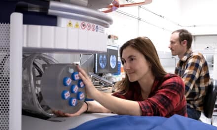
“For this new study, we developed a small diameter intravascular catheter that incorporates an optical fiber that delivers light to the coronary artery wall,” says Nadkarni. “We also used a small-diameter fiber bundle, polarizer and GRIN lens to image the reflected speckle patterns onto a CMOS sensor.”
For preclinical testing, the researchers evaluated the ability of their ILSI instrument to detect unstable plaques in a human coronary to swine xenograft model. This model system uses human coronary arteries that are sutured onto the beating heart of an anesthetized living pig. They assessed the mechanical properties of plaque inside the arteries by calculating the rate, or time constant, of fluctuations in the intensity of the speckle pattern and then compared their results with histopathological findings.
“The time constants in unstable plaques were significantly and distinctly lower than other stable plaques in the coronary wall,” says Nadkarni. “These results demonstrated the exquisite diagnostic sensitivity and specificity of ILSI for detecting human lipid pool plaques that were most likely to rupture under physiological conditions.”
The researchers say that the new technique could be easily integrated with other intracoronary technologies such as optical coherence tomography or intravascular ultrasound to combine the mechanical findings from ILSI with morphological information to improve the evaluation of plaque stability.
The researchers plan to continue to evaluate the capability of their ILSI instrument for rapid assessment of the coronary vasculature in live animals. Once these preclinical studies are complete, they will assess the safety of the catheter for use in humans and then begin the process of gaining regulatory approval for clinical use.