Summary: A study published in AJR found that machine learning radiomic models using multiparametric MRI showed suboptimal performance in noninvasively characterizing breast cancer by HER2 expression status, with SHapley Additive exPlanation analysis highlighting the importance of early-phase dynamic contrast-enhanced imaging and T2-weighted imaging features.
Key Takeaways
- The multiparametric MRI (mpMRI) machine learning models showed suboptimal performance in noninvasively characterizing breast cancer by HER2 expression status (HER2-zero, HER2-low, HER2-positive).
- SHapley Additive exPlanation (SHAP) analysis was used to interpret the model’s predictions, with early-phase dynamic contrast-enhanced (DCE) imaging being the most influential factor for both tasks.
- Despite the moderate area under the curve (AUC) scores of 0.757 and 0.754, the study highlights the potential of SHAP analysis for presenting complex radiomics data in a more accessible way for future research.
——————————————————————————————————————————————————————————————
Findings from an accepted manuscript published in the American Journal of Roentgenology (AJR) indicate suboptimal performance of multiparametric MRI (mpMRI) machine learning radiomic models for noninvasive characterization of breast cancer with three-tiered HER2-expression status: HER2-zero, HER2-low, or HER2-positive.
“The study provides an example of the use of SHapley Additive exPlanation (SHAP) interpretation analysis to better understand predictions of imaging-based machine learning models,” writes corresponding author Yuan Guo, MD, PhD, from the radiology department at Guangzhou First People’s Hospital in Guangdong, China. “The visual presentations from global and local SHAP analyses may provide a template for presenting complex data from future radiomics investigations in an accessible format.”
Guo and colleagues’ study, published in AJR, involved 737 breast cancer patients (average age 54.1) from two centers (center 1: 578 patients; center 2: 159 patients) who underwent breast MRI and HER2 testing after biopsy. The research focused on two main goals: (1) distinguishing HER2-negative (HER2-zero or HER2-low) from HER2-positive tumors, and (2) further separating HER2-zero from HER2-low tumors.
Patients from center 1 were divided into training (70%) and internal test sets (30%) for both tasks, with 405 patients used for training in task 1 (284 for task 2), and 173 for testing in task 1 (122 for task 2). All 159 patients from center 2 were used as the external test set. The researchers analyzed radiomics features extracted from dynamic contrast-enhanced images, T2-weighted images (T2WI), and diffusion-weighted images (DWI).
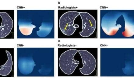
In external testing, the mpMRI radiomics score achieved an area under the curve of 0.757 for distinguishing HER2-negative from HER2-positive tumors, and 0.754 for differentiating HER2-zero from HER2-low tumors. SHAP analysis showed that early-phase DCE features had the strongest influence in both tasks, while T2WI features also played a significant role in distinguishing HER2-zero from HER2-low tumors.
Featured image: DCE, dynamic contrast-enhanced; T2WI, T2-weighted imaging; task 1, to differentiate HER2-positive from HER2-negative negative (i.e., HER2-zero or HER2-low) tumors; task 2, to differentiate HER2-low from HER2-zero tumors; SHAP, SHapley Additive exPlanation.