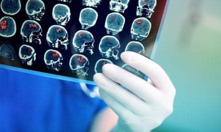
CT is one of the most powerful and well-established diagnostic tools available to modern medicine. An increasing number of people have been opting for CT scans, raising concerns about the amount of x-ray radiation that patients are exposed to. Ideally, a patient is exposed to minimum radiation levels during treatments or diagnostic procedures, while still receiving the expected benefit.
In practice, this is known as the ALARA principle, which stands for “As Low as Reasonably Achievable.” However, this principle results in a trade-off because CT image quality decreases with a decrease in radiation power. Thus, medical staff usually aim to strike a balance between a patient’s exposure to x-rays and obtaining good quality CT images to avoid misdiagnosis.
This balance can be achieved through an optimization strategy, in which healthcare professionals, primarily radiologists, observe real images generated by the tomographer and try to identify features, such as tumors or abnormal tissue. Following this, a specialist employs statistical methods to calculate the optimal radiation dose and configuration of the tomographer.
This procedure can be generalized by employing reference CT images obtained by scanning specifically designed phantoms containing inserts of different sizes and contrasts, which represent standardized abnormalities. Nevertheless, such manual image analyses are very time-consuming.
To address this issue, a team of researchers from Italy led by Sandra Doria, MD, and members of the Physics Department at the University of Florence, in collaboration with radiologists and medical physicists from Florence Hospital, explored the possibility of automating this process using artificial intelligence (AI).
As reported in Journal of Medical Imaging (JMI), the team created and trained an algorithm—a “model observer”—based on convolutional neural networks (CNNs), which could analyze the standardized abnormalities in CT images just as well as a professional.
To do so, the team had to generate enough training and testing data for the model. Thirty healthcare professionals visually examined 1000 CT images, each consisting in a phantom that mimics human tissue. Aptly termed “phantom,” this material contained cylindrical inserts of different diameters and contrasts.
The observers were asked to identify if and where the inserted object appeared in each of the images and state how confident they were in their assessment. This resulted in a dataset of 30,000 labeled CT images taken using different tomographic reconstruction configurations, accurately reflecting human interpretation.
Next, the team implemented two AI models based on different architectures—UNet and MobileNetV2. They modified the base design of these architectures to enable them to perform both classification (“Is there an unusual object in the CT image?”) and localization (“Where is the unusual object?”). Then, they trained and tested the models using images from the dataset.
Through statistical analyses, the research team evaluated various performance metrics to verify that the model observers could accurately emulate how a human would assess the CT images of the phantom.
“Our results were very promising, as both trained models performed remarkably well and achieved an absolute percentage error of less than 5%. This indicated that the models could identify the object inserted in the phantom with similar accuracy and confidence as a human professional, for almost all reconstruction configurations and abnormalities sizes and contrasts,” says Doria.
Doria and her team believe that with additional efforts, their model could become a viable strategy to automatically assess CT image quality. She adds, “Our CNN-based model observers could greatly simplify the process of optimizing the radiation dose used in CT protocols, thereby minimizing health risks to the patient, and help avoid the time-consuming limitations of medical evaluations.”
Doria anticipates that the team will succeed in applying their AI model observers on a larger scale, making CT evaluations faster and safer than ever before.


