Sharks, found in deep and shallow waters throughout the oceans, are some of the oldest living creatures on planet Earth. Shark skeletons, made of rubbery tissue called cartilage, have fascinated scientists for eons. Sharks swim at high speeds under deep water, and their skeletons experience high pressure and strain. These swimming-induced loads are borne by the centrum—mineralized bony tissue present in the shark’s vertebrae. However, from a functional standpoint, it is not fully clear how the complex 3D mineral structures of the shark centrum support and distribute loads within it.
To gain further insights into the special characteristics of shark centra, an interdisciplinary team of researchers used a novel approach in which energy dispersive diffraction (EDD) was performed using polychromatic synchrotron x-radiation. Their findings are reported in a new paper published in SPIE’s Journal of Medical Imaging.
“Studies that use 3D diffraction to map mineralized tissue are quite rare, and there have been no such diffraction studies on shark centrum tissues. The information we can get from traditional absorption or phase contrast imaging techniques is quite limited,” explains corresponding author Stuart R. Stock, PhD, of Northeastern University’s Feinberg School of Medicine in Chicago. “The mechanical properties of mineralized tissues depend strongly on how the mineral is oriented, and we wanted to use our novel technique to obtain informative 3D EDD maps of distribution of bioapatite nanocrystal orientations within the mineralized tissue of blue sharks.”
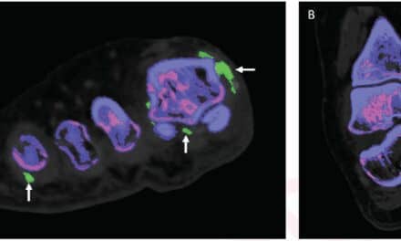
Unlike conventional x-ray diffraction techniques that use x-rays of one energy, EDD irradiates with x-rays of different wavelengths. Differently oriented nanocrystals diffract different wavelengths, so moving the specimen across the beam allows the varied crystal orientations to be mapped. The team combined the diffraction technique with microcomputed tomography and fluorescence mapping to add complementary information. The combined technique effectively allowed the researchers to see “inside” the centrum tissue at a molecular level and construct high-resolution 3D maps of the bioapatite arrangements within it.
The main structures of the blue shark centrum are the cone walls and wedges. The researchers found that the orientation of bioapatite differed between the cone walls and wedges and interpreted this as allowing the shark vertebra to resist lateral and axial deflections, respectively, during swimming. These findings offer interesting insights into the structure-function relationship of the shark skeleton and could perhaps be applied to bony structures and tissues in other organisms.
According to Bert Müller, medical faculty at the Switzerland-based University of Basel, “The combination of diffraction and tomography with fluorescence imaging data provides detailed insight into the crystalline organization of bone and cartilage down to the molecular level. It offers a starting point for a deep understanding of the unique oriented structures in bony tissues and the related skeletal function.”
By showing that mineralized tissue samples can be mapped three-dimensionally using EDD tomography, the study provides a proof-of-concept that has important implications for studying bones of human and animal cadavers in the medical sciences.
Stock says, “3D mapping with EDD could help us understand nanoscopic differences in mineral structure between healthy and diseased bones, as in osteoporosis, and also understand how healed bones are different from native bones. Such studies could lead to significant clinical advancements.”