By Fred Vernacchia, MD
In the past, many physicians have readily used dual-energy x-ray absorptiometry (DXA) as a convenient and reimbursable technique for assessing bone mineral density (BMD) in patients at risk of osteoporosis. However, there are severe limitations of DXA in measuring BMD in obese or arthritic patients. Clinical factors like these, coupled with the cut in DXA reimbursement (March 2012), has left practice managers and physicians looking for alternative BMD testing solutions that offer quality results across a wide range of patient requirements.
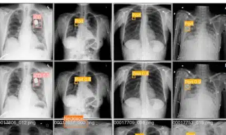
In this new market environment, the maturity of QCT technology and its adherence to low-dose CT protocols have propelled it to the forefront. QCT is a clinically effective BMD solution that offers better sensitivity to early changes in bone mineral density and, as a result, can allow for more effective patient management. In addition, the new iterative CT reconstruction methods being introduced by all the major CT manufacturers promise to reduce x-ray dose further and make QCT an even more attractive solution.
QCT at a Glance
QCT is a fast, noninvasive technology that performs BMD exams of the lumbar spine and proximal femur utilizing low-dose scan protocols on a standard CT scanner.
Originally developed in the early 1980s, QCT has several advantages over dual-energy x-ray absorptiometry. While DXA offered the industry a quick, low-dose option for performing bone densitometry exams, the downside was that DXA measurements were compromised by a number of common clinical conditions including obesity, scoliosis, disc space narrowing or spinal degenerative diseases, aortic calcification, and osteophytes in arthritic patients.
Despite these limitations, wide adoption of DXa over QCT was largely due to the limited availability and cost of access to early CT scanner time. In the last decade, however, scanners have become not only faster but also more widely available for conducting BMD exams. Furthermore, QCT of the spine has evolved from 2D scanning (using gantry tilt) to fully 3D volumetric scanning and has enhanced its ability to measure BMD quickly, efficiently, and with little user interaction, enabling excellent reproducibility.
Today, QCT is widely used for both routine and specialist BMD measurement in part because of the introduction of DXA-equivalent T-score measurement at the hip and easy connection to modern PACS infrastructure, and in part because of its superior cost effectiveness particularly for rural hospitals and other low volume facilities.
How It Works
QCT software calibrates an existing CT scanner according to known BMD reference standards. A typical QCT exam takes about 5 minutes or existing (non-IV contrast) abdominal/pelvic scans (ie, virtual colonography) can be used without the need to acquire additional images and expose the patient to any extra radiation dose.
Misconceptions
A number of misconceptions exist about QCT technology that have deterred practices from adopting QCT for BMD testing. Many believe that QCT is unsafe because it uses a higher dose of radiation. While QCT technology requires a higher dose than DXA, the radiation dose is comparable to other standard screening exams such as mammograms. Modern CT scanners make it possible to use low-dose image techniques, which means that QCT images can now be acquired with approximately one fourth of the radiation normally used for an abdominal or pelvic scan.
Misconceptions about the reproducibility of QCT also have resulted in the myth that QCT measurements are not as precise as those of DXA. This relates back to the early stages of QCT development, when older 2D QCT methodologies depended to a greater extent on the skill of the operator for BMD measurement. Modern QCT uses fully 3D volume scans, and bone density analysis is now largely automated.
In terms of precision, modern 3D QCT is highly reproducible with spine or DXA-equivalent hip measurement errors of around 1%, which is about the same as those from use of a DXA machine. But it is the differences between QCT and DXA that have a huge impact on the patient experience and treatment.
QCT’s 3D bone density capabilities allow it to make highly sensitive measurements of the metabolically active trabecular interior bone. Because trabecular bone loss or gain is commonly eight times more rapid than in the dense cortical bone that forms the exterior bone walls, QCT is likely to detect low bone mass in the spine earlier than other bone densitometry technologies and, as a result, facilitate earlier therapeutic intervention.
This ability to isolate and measure the trabecular bone independently also avoids the artificially high BMD measurements that often influence other DXA results and are due to obesity, disc space narrowing or spinal degenerative diseases, or aortic calcification and osteophytes in arthritic patients.
Treating physicians will recognize how these complexities can easily have an impact on a patient’s care, and using QCT technology has the additional benefit of being clinically time-efficient. Helical CT scans are completed within seconds, and an automated analysis of the exam is delivered within 5 minutes.
Clinical efficiency is further enhanced because the CT machine can scan in 3D, and a patient’s anatomy can be positioned after the scan using QCT software. This means that, unlike the positioning requirements of other technologies, the hips do not need to be rotated for a QCT scan, making a difference to patients with arthritic hips who may find rotation uncomfortable. Enhanced clinical utility also extends to spine BMD exams using QCT for patients with scoliosis and other spinal complications and in oncology patients who are prone to rapid and significant bone loss due to prolonged steroid or hormone therapy.
As a long-time user of QCT technology, I can testify to its flexibility to suit the needs of my practice. QCT scans and bone density measurements are performed by a CT technologist who is guided through the simple workflow by the software to produce a BMD report within minutes. Not only is most of the image analysis automated but also if a patient already has a historical report present in the database, it is automatically compared to the new results.
The dramatic cut in reimbursement for DXA in March 2012, with another cut likely in 2013, will support the more widespread adoption of QCT. QCT is reimbursable for routine BMD screening of a postmenopausal woman every 24 months. Other eligible beneficiaries for QCT BMD exams include patients with vertebral fracture or hyperparathyroidism, or those on long-term steroid therapy.
With no need for a dedicated technician or space as is required for a DXA machine, QCT can deliver a clear economic advantage.
###
Fred Vernacchia, MD, is a radiologist and founder of San Luis Diagnostic Center in San Luis Obispo, Calif. He has been using QCT technology since 1994 as an integral part of the state-of-the-art services provided at the center.