Radiologic technologists who are new to a digital facility or have left the field for a few years?say, to have a child?might wonder if they’re in the right place. Large consoles resembling elaborate video games loom in the peripheries of the workspace. Modern, extra-light cassettes abound. In some rooms, and in some entire imaging departments, cassettes are missing altogether. And that smell?that aromatic blend of fixer and developer that automatically pegs the locale?is noticeably absent. So is its source: the darkroom, the one-time mainstay of the department. It’s now a storage closet or someone’s office, complete with commonplace lighting. Like the black lights of the 1960s, the moody red glow of the safety light now appears relegated to a bygone era.
Welcome to medical imaging in the 21st century, a revamped and highly digitized experience. Perhaps the surprise is that it took this long. Such modalities as CT and MRI were digital beings at birth, but?until recently?they were required to yield “dumbed-down” film outputs to accommodate the lagging world of conventional X-ray equipment, their analog brethren. Now, finally, most major imaging departments have transitioned away from film for all modalities and embraced the great benefit of digitally stored images. So, amid all of these changes, where’s a new, transferred, or reinstated technologist?feeling not so brave in this modern world?to start? Right here, with a primer.
DR, Meet CR
Technologists generally will encounter three digitally oriented environments: strictly CR, strictly DR, or a combination of the two, according to Jacqueline Gallet, clinical studies manager with Eastman Kodak Co’s Health Group (Rochester, NY). CR incorporates familiar standbys, including a typical X-ray tube, a cassette, a table or stand, and a bucky or grid to help clean up the image. Rather than carting an exposed cassette to a darkroom, however, the tech inserts it into an imaging reader after keying in the type of procedure. The reader extracts an imaging plate from the cassette, and the image?which is read by laser lights?assembles on a monitor. In the meantime, the imaging plate is erased, and the cassette is ejected and ready for the next exam.
If the image passes muster, the tech generally sends it to the PACS, allowing a radiologist or other physician to access and manipulate it from virtually any location. While the image stores here in digital form, analog elements mark the CR process; hence, the system is not technically considered fully digital.
Although CR (teamed with PACS) eliminates the workflow associated with the film library, the system?in comparison to film-screen methods?will pare little time off the exam itself, Gallet says. “The steps in the front-end process of CR?placing the cassette in the bucky, removing it, placing it in the reader?aren’t that different from an analog film environment, with a central reader simply replacing the darkroom,” she says.
The biggest flaw associated with a darkroom or central reader is bottlenecking. To counter this issue, manufacturers like Kodak offer readers for individual workstations as well, Gallet adds. Some readers accept just one cassette, and others have multiple sleeves; regardless, image production is far from instant, as processing occurs one cassette at a time.
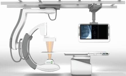
Technologists working with a DR system, in contrast, enjoy cassette-free environs. DR systems, which supplant any existing equipment, include digital-imaging sensors permanently mounted beneath an X-ray table or stand. After an exposure is made, the image assembles on a monitor at the workstation and, if acceptable, is whisked off to PACS in seconds without further ado. For streamlined workflow and efficiency, DR is unsurpassed, according to Gallet.
PACS?which nearly always accompanies a CR or DR system?remains the master link in the digital chain, notes Russell L. Cain, director of imaging at Haywood Regional Medical Center (Clyde, NC) and a continuing education instructor at the Medical Technology Management Institute. 1 “Facilities that have gone from film to digital or are making or planning the transition are pretty much driven by the move to PACS,” he says.
The evolution hardly happened overnight, adds George Spahn, director of image quality for FUJIFILM Medical Systems USA (FMSU of Stamford, Conn). “It was very difficult to displace film, because people were very comfortable with it. Once all of the advantages of PACS surfaced?the ability to manipulate the image and the accessibility of that image?the move toward digital systems really began to explode,” he says.
Cain says that although geography also plays a role, in general, the bigger the facility, the greater the chance a tech will see a PACS and a CR or DR system to support it. “[In the United States,] eight out of ten of large tertiary facilities with more than 500 beds have PACS, and many of those that don’t are making plans for implementation. For facilities with less than a hundred beds, as low as 26 percent use PACS,” he mentions.
Cain also notes, “While DR cuts a third off the time of processing and workflow, it is quite a bit more expensive than CR. Low-volume hospitals nearly always will go a CR route, as it enables the leap to digital and PACS at the lowest cost. Efficiency in these facilities is not as much of an issue.”
Kathy Pitura, research specialist for FMSU, agrees. “For facilities that already have made a recent investment in [standard] equipment, or simply don’t have an excess of capital funds, CR offers a tremendous amount of flexibility by allowing entire departments to go digital at a cost that’s significantly lower [than DR].” Still, DR’s appeal, and its implementation, is increasing exponentially, including with facilities that already have committed to CR. “Today, you’re quite likely to see a combination of CR and DR, as DR becomes incorporated, equipment is replaced, and the department upgrades,” Pitura says.
Cain adds that well-managed imaging departments are using foresight to prevent recent purchases from becoming outmoded. “Our hospital, for example, currently has a CR reader in every room,” he explains. “If I begin upgrading rooms to DR, for the efficiency and enhanced image quality, I might move a reader to the emergency department and continue to do portables there with the CR, or maybe move one to the operating room or critical care unit to handle portables in those locations. That’s [typical of] a phase-in process that will probably occur down the line.”
The portability of cassettes?and the flexibility they offer the tech, especially regarding emergency-department, trauma, and bedridden patients?have practically been CR’s raison d’?tre. That versatility, however, now extends into the DR world, with the advent of portable sensor panels, according to Herman Krug, product specialist for Canon Medical Systems (Irvine, Calif). The cassette-sized panel includes a cable that connects to a modified portable machine that houses a computer and monitor. Following a portable run, the machine is plugged into a network jack, and the images are downloaded to PACS, Krug explains.
He adds that a portable DR system can make life easier for both technologists and physicians. “Many times, you’ll face doctors who are still on the floor after placing a line in the patient,” he says. “After shooting the X-ray, the image appears instantly on the monitor. While the monitor on the portable isn’t of diagnostic quality, it’s adequate to allow a doctor to decide whether a line needs adjusting. If it does, the tech can repeat the shot right on the spot.”
Cain opines that such a system might be a wise investment for deep-pocketed, large medical centers that manage a vast number of portables per day. However, he notes that the panels are expensive, are heavier than typical cassettes, and bear a fragility factor.
An Expos? on Exposures
 Both CR and DR systems offer the benefit of a much larger dynamic range, or latitude, than film/screens, and they accept a much greater range of exposures, according to FMSU’s Spahn. To the technologist, this means that the number of repeat examinations due to under- or overexposure will be reduced significantly. The compensating factors inherent in digital processing make for smooth transitions when technologists adjust their techniques, or exposure factors, from film to digital systems.
“The basic rules that govern imaging for film-screen systems are still in effect for digital systems,” Spahn says. “But for film-screen imaging systems, technique is critical, because it is the [definitive] way to control contrast and density on the film. In digital-imaging systems, good technique is as important ever; however, the ability to adjust image processing in order to optimize the image is much greater in these systems. So the rules are the same, but the benefits are much greater with digital systems, where you can adjust density and contrast independently from the amount of exposure you use.”
Film’s relatively unyielding medium cannot compete. Short-latitude film produces excellent skeletal images, but it offers poor views of the surrounding soft tissue. Wide-latitude film might present fine views of certain soft tissue, but it sacrifices an optimal look at the skeletal structures in the region. “In film-screen [environments], you have a fixed image?one presentation, which limits the amount of information available,” Spahn notes. “Using a hot light, for example, is an attempt to make the presentation more flexible. The digital domain offers significantly more flexibility for the radiologist to optimize the image. This adjustability is especially advantageous for areas like the chest that offer such a varying range of density.”
However, the wide dynamic range of the CR or DR system offers somewhat of a “dual-edged sword,” 2 according to the Sprawls Educational Foundation (SEF of Montreat, NC). Its plus is that, unlike with film, a varied range of exposures?including errant ones?will render films with good contrast. Two prominent downsides exist, however: the potential of either underexposures that can discreetly mar and limit information, or overexposures that have aroused new ethical issues in the field.
The Nature of Noise
According to the SEF, although inadequately low exposures can render images with good contrast, a high level of quantum noise will be present. An abundance of noise is a common adverse effect in the digital process and can render an image undiagnostic.
“Noise appears as graininess?the analogy is snow on an old television that receives a poor signal,” Canon’s Krug explains. “For radiographic procedures, the tech needs to determine the optimum technique that minimizes both image noise and patient exposure. All digital modalities, however, inherently have some degree of noise.”
That inherence can take some adjustment for those unfamiliar with digital outputs, Spahn notes. “Noise occurs in an area of the image that doesn’t receive enough information through that body part, so it isn’t uniform in an image. If you view a chest X-ray, for example, the lung fields?filled with air?have a lot of penetration and, thus, a good signal. With digital imaging, you can amplify weak signals, like through the heart and mediastinum?areas that were basically white [and devoid of information] on film?but this will result in a graininess or loss of smoothness, which can take a little getting used to for those accustomed to film.”
Spahn adds that while upping the technique in his example isn’t necessarily a beneficial way to reduce a slight graininess of the mediastinum, most customers simply need time to acclimate to the distinctiveness of the medium.
Overexposures: Easy on the Trigger
Unlike the underexposed film, the overexposed image on a CR or DR system is of very good quality, according to the SEF. And although radiologists no longer have to contend with a dark film masking information, the negative consequence is that it is easy for technologists, especially those unsure of their techniques, to expose patients to excessive radiation.
“The philosophy [in medical imaging] has always been to use the minimal amount of dose necessary in order to get the level of image quality that is necessary,” Spahn says. “Techs know, however, that if the digital image is overexposed and the system corrects for it, the image quality is never going to come into question. Yet, it’s done at a level of exposure that is unnecessary to the patient.”
Ironically, overexposure lacks any value whatsoever in the digital realm. “In the film world,” Spahn says, “you might need to overpenetrate the chest, for example, to accentuate the placement of a line. In the digital realm, that’s no longer necessary, because you’re able to bring out that detail through the image processing.”
Digital systems offer a method of monitoring exposure levels by producing a numerical output following an exposure, explains Haywood Regional’s Cain. Low exposures render a high calculated number (for example, S=400), and high exposures produce a low number (S=50). Ideally, this number should fall in an exam-specific numerical range generically called the sensitivity index (also known as proprietary-specific terms like “S Number,” “Rex Number,” “exposure index,” etc). This quantitative method is the optimal way to gauge exposure levels, as the brightness and contrast of the image provide few clues in the digital world, Cain notes.
“Fuji’s S-value limits are actually designated by the facility for different examinations,” Spahn says. “If an exposure is outside of those ranges, the technologist receives a notification.” Adds Penny Maier, marketing manager for FMSU, “It is possible to generate a report showing the S values for all exams during a designated period of time. That data can be sorted easily to identify exams that were out of recommended range.”
Ultimately, however, it is the facility’s decision whether to implement policies that address and monitor patient exposure.
Opening Windows
Even though an overexposed exam will yield a very good X-ray, it will not equal an optimally exposed image, Cain says. “What you lose?if you’re off [either side of] the S number?is the full range of the ability to window and level the image.”
Spahn adds, “Window width and level?the ability to adjust density and contrast of the image?is a valuable tool for diagnosis. However, we recommend that the technologist not adjust the image’s window width or level [prior to sending it to PACS]. If the image doesn’t look as well as it should, the processing parameters should be changed to optimize the image instead. When a tech windows and levels, the data that goes out to PACS is actually truncated, and the physicians have less capability to work with the image. So it’s important that the images are sent out at full window width and contain all the diagnostic information.”
As for technologists who are embarking on digital imaging but are considering the window to be half-closed rather than half-open? Once they start working with the technology, they’ll realize the similarities to film more than the differences, Spahn says.
“What makes a technologist a good technologist are still the same exact things, whether it’s the film-screen world or a digital world,” he says. “Although digital systems are more forgiving in variances in technique, the fact is that a poorly positioned exam will be repeated regardless of systems. The skills don’t change, but some new skills are required as well, including understanding computer systems as well as aspects of image processing to ensure optimal image quality. Applying what you’ve learned in the past and continuing your education are as important today as ever.”
Mark Dye, RT, is a contributing writer for Medical Imaging.
References
- Medical Technology Management Institute. The evolution to all- digital radiography. Available at: www.mtmi.net/seminars/ evolution.php . Accessed March 7, 2006.
- Sprawls Education Foundation. The digital radiography system. Available at: www.sprawls.org/resources/DIGRAD/ module.htm#1 . Accessed March 7, 2006.