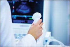
 From Top to bottom: Holigic’s new dexa discovery, GEMS’ lunar’s prodigy vision, The excell plus from cooper surgical
Often called the “silent disease” because bone loss occurs without symptoms, osteoporosis poses a significant public health threat for adults in the U.S. and around the world.
According to the National Osteoporosis Foundation (Washington, D.C.), an estimated 10 million individuals in this country exhibit hallmarks of this disease, with almost 34 million more showing evidence of low bone mass placing them at increased risk for developing osteoporosis. Eighty percent of those affected by osteoporosis are women. NOF estimates that national direct expenditures for osteoporotic and associated fractures was $17 billion in 2001 ($47 million each day), with costs increasing as healthcare expenses rise.
Nelson Watts, M.D., director of the University of Cincinnati Bone Health and Osteoporosis Center (Ohio) explains the importance of screening for the disease in all women 65 and older to determine patients at risk of developing the disease. The NOF includes women who are less than 65, but who meet certain risk criteria such as a personal history of fracture after age 50, a family history of osteoporosis, current cigarette smoking, or estrogen deficiency as a result of menopause, especially if it occurs early or is surgically induced.
Although treatment is available, in order to take advantage of the new drugs that can prevent onset of debilitating fractures, early detection is required. An emphasis on screening with bone densitometry has served as the vanguard for the initial diagnosis of persons who fall into high-risk categories.
Laura Tosi, M.D., chair of women’s health issues for the American Academy of Orthopedic Surgeons, and chief of orthopedics at the Children’s National Medical Center (Washington, D.C.) proposes a paradigm shift in the management of patients with fractures of the hip, wrist or ankle. Citing significant data that reveal that if a woman breaks her wrist in the perimenopausal period, there is increased likelihood that she will fracture her hip at a later date, or a person with one vertebral compression fracture is at increased risk of suffering a similar fracture in another vertebra, Tosi suggests that orthopedists could help the situation by recognizing adult fragility fractures as a possible indicator of osteoporosis.
“There is a growing body of data that indicate that if you fracture as an adult, not even post-menopausal, just a man or woman over 45 to 50, particularly if you have a fragility fracture, you are somebody who is likely to re-fracture,” says Tosi. A fragility fracture is defined as a low energy fracture resulting from a fall of standing height or less.
With 1.5 million fractures per year in the United States, and a high percentage of those considered fragility fractures, Tosi proposes that all of those individuals deserve a workup to rule out some underlying health problem.
“Somewhere between 30 percent and 60 percent of people with osteoporosis have an underlying treatable condition,” continues Tosi. “It might be hypothyroidism, hypogonadism if you’re a man, calcium wasting by your kidneys, but there may be something we could treat you for today.”
For the past several years, treatment has included the administration of oral bisphosphonates such as Fosamax (Merck & Co., Inc) or Actonel (Procter & Gamble Pharmaceuticals) — considered antiresorptive drugs that slow or stop bone loss. In December 2002, the U.S. Food and Drug Administration (FDA) approved the use of parathyroid hormone (PTH), with a drug called Forteo (Eli Lilly and Company) that is the first in a new class of drugs called bone formation agents. This drug stimulates bone-creating cells to produce new bone.
Testing … One, two, three
Bone Densitometry measures the mineral content of bones for both the diagnosis of bone loss, or to follow the response to treatment for those individuals who take medication.
“I think that for now and the foreseeable future, central DEXA (dual energy x-ray absorptiometry) measurements of the spine and hip will remain the gold standard both for diagnosis of osteoporosis and for monitoring treatment,” says Watts who serves as immediate past president of the International Society for Clinical Densitometry (ISCD).
Although peripheral tests are very useful for fracture risk assessment, there are some problems with employing these devices for screening, according to Watts. The first problem is that there is great variability with the different devices. The second is determining the exact “cut point” number to make results valid within the context of the WHO definition of osteopenia and osteoporosis.
Watts describes two patients who illustrate the difficulties. One was a 59-year-old woman whose ultrasound heel measurement showed her with a T score of minus 1.9, but her central DEXA was normal. The other patient was a woman who had received a normal result from a heel study, whom he saw the next week with a broken hip.
One of the issues with ultrasound heel devices involves accurate positioning of the foot to ensure that the correct bone is being measured.
Terri Bresenham, president and general manager of GE Lunar (Madison, Wis.), agrees that not all devices are created equal. She explains advances in the Achilles InSight that address the issue of appropriate scanning.
One of the breakthroughs with this device is the capability of producing a real time image of the heel. When the patient’s foot is placed, they can immediately see the calcaneous bone to ensure correct alignment. The second advance is that they have developed a gel-free method (using an evaporating spray) of scanning that proves helpful for screening large numbers of people.
 GEMS Lunar’s Prodigy Vision uses dual-energy assessment of vertebral bodies to better detect faractures.
Central DEXA, still the gold standard bone density testing method using DEXA technology has served as the basis for the WHO standard definitions of osteopenia (bone loss) and osteoporosis.
“The peak bone density number is 180 mg, based on calcium hydroxyapatite which is a mineral salt that makes bone hard,” explains Steven Knox, chief technologist, Health Perfect Imaging (Costa Mesa, Calif.).
If a bone mineral test shows 29 milligrams less of this mineral than is normal for a 30-year-old woman (or man), a T score of minus one is assigned because the reading is one standard deviation below normal. Osteopenia is defined as a T score of minus 1 to minus 2.5, and osteoporosis is defined by WHO as a T score of 2.5 (or more) standard deviations below normal.
Dual energy x-ray absorptiometry is a study completed by measuring the amount of x-rays that are absorbed by the bones in the body. Two x-ray energies allow the machine to differentiate between bone and soft tissue, to provide an estimation of bone density. Central DEXA is completed with studies of spinal bones in the lumbar region, and the hip.
GE Lunar’s Bresenham explains recent advances to their central DEXA product: Prodigy Vision with narrow-angle fan beam technology. The digital acquisition of data leads to precision in measurements and the dual-femur capability allows studies of both hips.
“About 25 percent of people have a significant difference between their two femurs,” says Bresenham. By measuring a larger portion of the body, more statistics are generated, leading to greater precision. This capability is also important when performing longitudinal studies over the years in the event a person fractured one hip. If that were the femur being followed, obvious problems would arise, so it becomes a precautionary measure to study both hip bones.
The other reason that dual hip measurements prove valuable is that the spine becomes more difficult to accurately assess as a person ages because natural deformations occur over time.
Another advance application available on the Prodigy Vision is dual energy assessment of vertebral bodies, where soft tissue is “subtracted away” to enable visualization of the outline of the vertebral bodies that improves the ability to detect fractures. Computer Aided Densitometry is a feature that enhances accuracy during performance of examinations to eliminate variability from one technician to another in the manner they perform exams.
Hologic Inc. in Bedford (Mass.) has introduced their latest central DEXA product called Discovery that reduces scan time by 66 percent, providing a 10-second scan of the hip and spine.
 Mindways Software Inc.’s QCT Pro Bone Density software.
“The Discovery is unique because it combines not only a 10-second bone density analysis, but also has the ability to perform a 10-second image of the lumbar and thoracic spine in both the AP and lateral views,” explains Hologic’s senior product manager of the osteoporosis assessment division, John Jenkins. This feature, called Instant Vertebral Assessment (IVA) is a rapid imaging technique designed to evaluate spinal fractures at the point of care.
The Discovery system provides digital acquisition of images and data, enabling DICOM image integration to worklists, remote computers or PACS. This equipment is designed to improve throughput and save time both for scanning and reporting functions.
Norland, a Cooper Surgical Co. (Trumbull, Conn.) has been involved in producing bone densitometry systems for the past 50 years. In April 2002, Norland was acquired by Cooper Surgical.
“Our prime interest has always been on offering the most economical bone densitometry systems, both central and peripheral to offer the precision and performance of our competitors … at a price point that is feasible for the primary-care physician,” says Norland’s vice president of worldwide sales and marketing, Marc Zimmer.
Norland offers two central DEXA products: the Norland XL that has the smallest footprint of any of the central units. With a six-foot table, this system can fit into a room that is seven foot by seven foot, and is capable of performing hip, spine and forearm measurements.
Their top-of-the-line offering is the Norland XR-46 that uses pencil beam technology and an eight-foot table to capture data over one and a half minutes to 5 minutes for the entire body. The fracture risk assessment feature provides simplified color reporting.
QCT to the fore
Although QCT has been available for many years, this technique applied to bone mineral density testing has gained enthusiasm from a number of practitioners.
Driving forces include the huge installed base of CT scanners, including fast multislice capabilities. With the addition of specialized computer software, an institution can use their CT to perform BMD studies and thereby generate increased revenue. Volumetric measurements enabled by CT are another factor in the plus column for the use of QCT for these studies.
The downside of using QCT is the capital expenditure for a CT scanner as opposed to a DEXA system, but if an institution already had a CT scanner in place, the economics might prove appealing for the use of this technique. Another disadvantage is CT’s higher radiation dose.
Jean M. Weigert, M.D., director of women’s imaging for Mandell and Blau M.D.s, PC (New Britain, Conn.) describes herself as an advocate for the use of QCT. Using Mindways Software Inc. (San Francisco, Calif.) QCT Pro with their GE LightSpeed CT scanner to perform both hip and spine assessments, Weigert explains that there are several circumstances where QCT provides more accurate measurements than DEXA.
“DEXA measures all of the bone: cortical, trabecular, etc. and summates the entire bone,” says Weigert. “So if you have arthritis with abnormal additional bone it will look like you have a normal bone density or high bone density.” With QCT, only trabecular bone, which is the most metabolically active portion of the bone, is analyzed.
In addition to those degenerative changes that occur in older patients, many in that age group demonstrate additional calcium in the aorta which lies right on top of the spine. Weigert notes that in those cases, DEXA will superimpose density from those calcium deposits onto the final reading. QCT will not.
Finally, Weigert prefers QCT because it is a volumetric measurement as opposed to an area measurement employed by DEXA.
“You’re taking a cube of bone from the center of the vertebral body, so it’s independent of the size of the bone,” says Weigert. “There’s always been the notion that short people have a higher risk of osteoporosis.” She believes it may be a factor of having smaller vertebral bodies. Volumetric measurements compensate for the variability.
Pat Feuerriegel, Mindway’s sales manager says that with QCT, changes in BMD are visible two to four times faster than with DEXA because in the spine QCT measures only trabecular, or metabolically active bone. The software is compatible with any CT scanner capable of producing DICOM images.
Mindways has just introduced a newly FDA approved hip module called CTXA Hip for including bilateral analysis of hip bones. Useful in situations where a whole-body scan is done, images can be evaluated for BMD. This system employs a phantom to calibrate the system to each scanner.
Joel Bortz, F.R.C.R., CT radiologist for Health Scan Imaging in Palm Desert (Calif.) used DEXA for many years, but now employs QCT software from Image Analysis Inc. (Columbia, Ky.) on their Philips Medical Systems Mx 8000 multislice CT scanner.
“Probably the biggest downside for DEXA is if you have degenerative disease in your spine,” explains Bortz. “If you’re doing QCT, you only measure the inside of the vertebral body. You put the marker where it needs to measure, and thereby exclude all extraneous disease such as degenerative changes in the spine and calcium in the aorta.”
Knox at Health Perfect Imaging uses Image Analysis software with their GE Imatron C-300 electron beam scanner, which means there is a lower radiation dose than would be found with a typical CT.
Roger Schulte, vice president of sales and marketing for Image Analysis, explains that they are in the process of submitting a proposal for a hip analysis module to the FDA. One of the challenges to adoption of QCT is that all of the data generated from the large population studies that informed the WHO standards for definition of osteopenia and osteoporosis were accomplished using DEXA systems. Schulte notes that the comparative literature suggests that QCT is more accurate than DEXA.
Act now
Osteoporosis causes significant morbidity and mortality for the patients who develop this debilitating disease. Data show that when people know that they have a health issue such as bone loss, they are much more likely to take action to correct their condition such as increasing their calcium and vitamin D intake.
With advances in both prevention of worsening bone loss and promising treatment for actual bone formation, accurate assessment of bone health becomes vitally important to the management of this disease. Bone densitometry through either DEXA or QCT measurements provides valuable data for these patients.
Our aging population can benefit from this information now, and increasing numbers of people will need it in the future.


