The only constant in women’s healthcare is that technology is constantly changing. From better ways to image breasts to cutting-edge treatment for uterine fibroids, the simultaneous evolution of multiple imaging modalities holds great promise for today’s clinicians?and their patients.
Counting on CAD
Approved by the FDA for clinical use in 1998, CAD for mammography employs algorithms to review images from mammograms as well as flag areas that warrant additional attention from a radiologist. Though initially thought by many to be overly sensitive, today’s CAD systems are clinically more effective. Recent studies suggest that fewer false negatives occur if two radiologists interpret a mammogram and include the use of CAD as that second look.
“Much like a spell-checker, CAD reduces the errors that radiologists inevitably make simply because the job is overwhelming in terms of the volume of cases that need to be read and the level of attention that needs to be maintained,” says Scott Parr, president and CEO of iCAD (Nashua, NH). Parr adds that in studies where CAD was applied retrospectively, it highlighted up to 72% of missed cancers.1
This sensitivity is a welcome addition for busy radiologists. “I’m comforted in particular by its ability to detect microcalcifications, because at the end of a long day, it can be hard to make sure you picked those up,” admits Debra Mitchell, MD, radiologist and partner at Breast Imaging of Oklahoma (Edmond, Okla). “I can’t say it changes my practice dramatically, but I don’t want to read without CAD. Its ability to back me up is comforting.”
Despite its effectiveness, the automated review process is intended to provide a safety net for physicians, not replace them. Relying solely on CAD would not only lead to many missed cancers, it also would result in many unnecessary biopsies of benign lesions.
Dynamic Duo
 |
| The Second Look system from iCAD helps radiologists detect breast cancer earlier by using patented CAD algorithms to mark suspicious areas. |
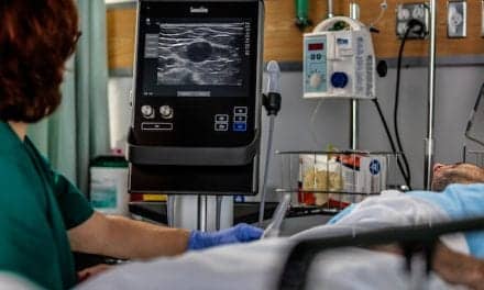
In general, ultrasound is strong in areas where mammography is weak. Specifically, mammography is best at detecting abnormalities in the fatty breast, while ultrasound excels in dense breasts, making them an ideal pair. “Ultrasound is a wonderful complement to mammo,” Mitchell says. “It’s not a case of one replacing the other; it’s that when you add them together, they do a great job.”
In addition to enabling radiologists to obtain quality images from different breast types, ultrasound also is used to further characterize things seen on a mammogram or something felt by the patient, but not visible with mammography.
“For example, if we see a mass on the mammogram, the ultrasound can tell us if it’s fluid-filled and benign, or solid and potentially malignant,” explains Constance D. Lehman, MD, PhD, director of breast imaging and associate professor of radiology at the University of Washington School of Medicine (Seattle). Lehman anticipates the day when manufacturers combine these two powerful modalities. Currently, performing both exams means moving the patient between machines, which is not only an inconvenience for her but is also something that can impact exam results.
“When a woman receives a mammogram, she is in one position. For an ultrasound, she’s in another, which means there’s a risk of missing the lesion,” Lehman explains. “Rolling these two technologies into one diagnostic tool would mean less likelihood of mismatching while making the information available at the same time.”
Magnetic Pull
Breast MRI is becoming a critical component in characterizing abnormalities initially identified through either mammography or ultrasound. The rapid increase in breast MRI use is due, in part, to the large amount of data demonstrating its strength as a tool to accurately detect, diagnose, and treat breast cancer, according to Lehman. For a myriad of reasons, breast MRI is not well-suited as a stand-alone screening modality. Not only is the exam expensive, but it falls short in many areas where the mammogram excels.
“We absolutely see breast MR as a complement to mammography,” Lehman emphasizes. “At this time, we know?without any question?that MR can find cancers that are hidden on mammo. But we also know that the mammo can identify cancers that are invisible to MR.” One example where mammography is generally superior to breast MR is in the identification of low-grade ductal carcinoma in situ.
Although it is not currently in demand for screening, breast MRI is highly valuable in other areas. For example, determining the extent of disease is often easier with MR than other modalities.
“We [scan with] MR all of our newly diagnosed breast cancers, and probably 10 to 15 percent of the time, we’ll find an additional area of malignancy?whether it’s fairly close to the original tumor or in the opposite breast,” Mitchell notes.
Research also indicates that some patients warrant breast MRI, including women diagnosed with an invasive lobular carcinoma, women with very dense breasts, or women who have ambiguous findings on mammography or ultrasound.
Breast MRI also can serve as a noninvasive, interim step between detecting an abnormality on a mammogram and performing a biopsy. The quality of an MRI helps radiologists determine if a mass is normal breast tissue or something that demands further investigation. And for patients undergoing neoadjuvant chemotherapy to shrink tumors before surgery, MRI can monitor the effectiveness of treatment and gauge the size of the tumor before and after chemo. Still, studies show there is room for improvement in this area. “Although MRI can be helpful in assessing response to chemotherapy in patients with advanced breast cancers, we also have found that MRI could underestimate the true extent of disease after the patient has received chemotherapy,” Lehman says.
As promising as it is, breast MR definitely has challenges to overcome, she admits, such as establishing industry-wide scanning protocols and interpretation guidelines. “It’s a challenging technology,” Lehman says, “and there’s quite a bit of variation to how breast MRs are performed and interpreted.”
Existing research demonstrating the positive role of breast MRI in breast-cancer detection and diagnosis was conducted at centers where the staff had some experience with the procedure, Lehman says. Whether or not those findings will transfer to facilities with less experience has yet to be determined.
The Guide to Biopsy
|
Using MRI to guide placement of a core needle, physicians are able to biopsy masses not visible with mammography or ultrasound. Though it is often a time-consuming process, using MR to perform biopsies relieves the frustration felt by many imagers who were early adopters of breast MRI as a diagnostic tool.
“When we first started doing breast MRI, we weren’t able to perform MR-guided biopsies,” says Kimberly K. Amrami, MD, chair of the division of body MRI at the Mayo Clinic (Rochester, Minn). “That meant, we’d see something we were worried about but then couldn’t find it on ultrasound or other tests.”
The turning point for Amrami’s team came after they acquired an FDA-cleared, vacuum-assisted, MR-compatible biopsy system from Suros Surgical Systems Inc (Indianapolis). “It’s very easy to use, and with the vacuum assistance, you don’t have to be exactly on the lesion,” she explains. “So you can be very confident that you’re actually biopsying the tissue you intended to biopsy.”
Image-guided biopsy also has a very high level of accuracy, a significantly lower cost, and a lower morbidity rate than traditional procedures for treating breast cancer.
A Full Field
In many areas of imaging, digital technology is replacing film with little effort. The transition has been a bit slower with mammography?and for good reason.
“The image quality is very good, and a lot of evidence shows that it is comparable to film-screen mammography,” Amrami says. “But there has not been any evidence to show it is better.”
Although workflow benefits of digital imaging include faster transmission, storage, and retrieval of images, the most important question has yet to be definitively answered. “What women really want to know is, ?Will it be better at detecting my cancer?,'” Lehman says. “And the jury really is still out on that.”
Fortunately, the wait is almost over. The American College of Radiology Imaging Network (ACRIN of Philadelphia) recently conducted an extensive trial comparing full-field digital mammography to film-screen mammography. Results from the Digital Mammographic Imaging Screening Trial (DMIST) were scheduled to be announced at the ACRIN Fall Meeting, held September 15?18 in Arlington, Va; however, they were not available at press time.
A Hot Focus
Not long ago, uterine fibroids were a common problem with few treatment options?but times have changed. Last fall, the FDA approved the ExAblate 2000, a system from InSightec Image Guided Treatment Ltd (Dallas) that uses MR-guided focused ultrasound (MRgFUS) for noninvasive treatment of uterine fibroids.
“Using MR guidance, we focus an ultrasound beam that heats the fibroid to a point where the tissue coagulates and is no longer viable,” Amrami explains. “The fibroids shrink over time, and patients have significant improvement in their symptoms without hysterectomy or myomectomy.”
The use of a highly focused beam means that concentrated energy from the ultrasound can pass through healthy tissue without damaging it. Analogous to the conical shape created when a magnifying glass is held in sunlight, only the focused point creates heat.
“We’re effectively doing the same thing with ultrasound as a source of energy rather than light,” says Paul Curtis, MD, interventional radiologist for Virtua Health (Voorhees, NJ) and South Jersey Radiology Associates. “It’s proved to be a very safe procedure with very few complications.” During clinical trials, only two patients of 109 experienced complications, he adds. One woman suffered a temporary nerve injury; the other had minor skin burns. Corrections to the processes were made to help avoid recurrence.
Although it has clear benefits, MRgFUS does place additional demands on the radiologist. Because the ultrasound beam is focused within a millimeter of accuracy, treatment progresses at a pace of about 1 cm3 per minute. Depending on the patient’s total tumor load, treatments could last up to 4 hours.
“Also, the radiologist must be present during the entire procedure, because in most cases, you have to make some kind of adjustment between every treatment point,” Curtis says.
As promising as it is, the treatment is not ideal for everyone. Women must be able to enter the MRI (eg, be free of pacemakers or aneurysm clips), be willing to lie prone for hours, and be motivated enough to stay alert and communicate with the physician throughout the procedure. “It’s critical to know if she feels anything in the sacrum or the sacral nerves, for example,” he says. “Depending on her feedback, you adjust the angle of the beam or change the energy.”
Additionally, the ExAblate 2000 is intended to treat only “women who have completed child bearing or do not intend to become pregnant,” according to the system’s FDA approval statement.2
Currently, almost 25 treatment centers worldwide offer MRgFUS, and additional sites are planned. At this time, the ExAblate system can be attached only to the standard 1.5T Signa MR scanner from GE Healthcare (Waukesha, Wis), though clinical trials are under way involving the company’s Excite 3T system. A study is taking place to determine the ExAblate’s ability to treat benign breast fibroadenomas.
Seek and Destroy
Uterine fibroid embolization, also known as uterine artery embolization (UAE), is a relatively recent addition to uterine fibroid therapy that involves routing a catheter into the pelvic region and through the arteries to deposit tiny particles that block the flow of blood to the fibroid.
Conducted by an interventional radiologist, this procedure shrinks the fibroid over time and might even prevent future growths. Most women with symptomatic fibroids are good candidates for UAE; however, it is not considered suitable for patients who want to become pregnant.
Both ultrasound and MRI are routinely used to diagnose fibroids, and, in many cases, an MRI usually is performed prior to UAE to determine their number, size, location, and enhancement pattern, according to Ajay Singh Sufi, MD, interventional radiologist at Community Radiology Associates (Rockville, Md). “The same technology also can help determine the effectiveness of therapy as well as assist in the evaluation of possible complications,” he says.
Connecting Technologies
Regardless of the modality in question, advancements in technology are only as good as the professionals they help. Exploiting and combining the strengths of individual systems will create a powerful diagnostic cache for physicians.
“We’re realizing these different imaging modalities are complementing each other,” Lehman says. “Our job is to match the imaging tools we have with each specific patient and the specific questions we have in that patient.”
Dana Hinesly is a contributing writer for Medical Imaging.
References
- iCAD. Second Look 6.0 Device Labeling. FDA approved PMA (Pre-Market Application) supplement for Second Look. October 31, 2003.
- FDA’s Center for Devices and Radiological Health: New Device Approval: ExAblate 2000 System ? P040003. November 3, 2004. Available at: www.fda.gov/cdrh/mda/docs/p040003.html. Accessed August 31, 2005.
Physiological Process Holds New Potential
Angiogenesis is the body’s process of creating new blood vessels from existing vessels. In healthy adults, it occurs primarily to heal wounds and as a natural part of a woman’s menstrual cycle. Otherwise, angiogenesis is seen only in disease. “More than 70 different diseases are angiogenic in nature,” says Phillip C. Thomas, CEO of DOBI [Dynamic Optical Breast Imaging] Medical International (Mahwah, NJ), “including rheumatoid arthritis, macular degeneration, and virtually all cancers.” Without a dedicated blood supply feeding it oxygen and nutrients, most tumors will not exceed 1 mm or 2 mm in diameter. Research indicates that most cancers are angiogenesis-dependent once they grow beyond approximately 5 mm. The vessels that form to feed a tumor often grow to be larger than the lesion itself, creating an area of vascularity significantly different from normal blood supply. “If we see vessels that are tree-like in shape, we know they are normal,” explains Michael Crade, MD, medical director of Ultrasonix Corp (Long Beach, Calif) and associate professor of radiology at University of California?Irvine. “But where there is angiogenesis, the arteries and veins are chaotic.” According to William W. Li, MD, president and medical director of the Angiogenesis Foundation (Cambridge, Mass), finding this disorderly group of vessels could allow clinicians to identify a cancerous lesion at its smallest size, possibly even before current modalities would be able to detect the tumor mass. “For diagnostic purposes, angiogenesis detection is likely going to play an adjunctive, not a replacement, role with other current technologies,” Li says. He adds that although several imaging modalities have been adapted to detect parameters reflecting tumor blood supply, “none of these technologies has yet been validated for accurately capturing angiogenesis in cancer.” One such system working toward FDA approval is manufactured by DOBI Medical International. The ComfortScan system is a stand-alone investigational device able to locate angiogenesis in breast tissue. “There’s generally a steady-state condition in the breasts that would allow for consistent use of the ComfortScan system in identifying abnormal neovascularization,” says Thomas, adding that the only known exception is expectant mothers. “During pregnancy, the blood supply to the breast is altered, which could mask angiogenesis associated with a lesion.” Thomas notes that systems like the ComfortScan will be most productive when used in concert with existing modalities. This combination of existing and cutting-edge technologies appears to be the future of medical imaging. “We see it being used to help further characterize a mass that mammography identifies,” he says. “If a patient has an indeterminate mammogram, a ComfortScan could be performed to help characterize whether the lesion is malignant or benign.” Unlike mammography, breast density is not as great an issue for the ComfortScan system, which is currently undergoing a large clinical study with FDA submission to follow its completion. But that’s not to say this innovative approach is on hold. Since acquiring a GE Healthcare 730 Voluson ultrasound system more than 1 year ago, Crade has changed the way he looks for ovarian cancer. “The system uses color-flow Doppler to capture the 3-D volume of whatever you’re looking at?in this case, the ovary,” he says. “But it not only captures the volume; it also captures the vascular data in 3-D. It’s truly revolutionary.” The ability to see vessels in brilliant reds and blues makes a world of difference, Crade says, because in 2-D black-and-white images, benign ovarian masses can appear malignant. “Something may look abnormal on regular 2-D and be suspect of cancer,” he explains. “But if, by using color, you can see the vascularity is normal, then you save a lot of people from unnecessary surgery. I think everybody wants to do a better job?this is one way to do it.” Crade and his team also employ the Voluson to characterize abnormalities in the uterus to determine, for example, if a mass is actually inside the endometrium or outside, pushing into the canal. Regardless of the organ in question, Crade believes the ability to see the vascularity in 3-D improves his diagnostic capabilities. “It’s given me a much greater confidence with diagnosis,” he says, noting that improved patient care is the bottom line. “The goal of the report is not just to say what’s on the films. The goal of the report is to say what the patient has and to be right about it.”
|
||||
?DH |


MAKING MAMMO MORE COMFORTABLE
There is little argument in the medical community that mammograms play an important role in breast-cancer screening. But studies suggest that many women neglect annual mammograms due, in part, to anxiety about the procedure. The rising popularity of devices created to increase a woman’s comfort during the exam?such as the MammoPad?might not only decrease a patient’s hesitation but net better images in the process. “We conducted a trial study to see if using the MammoPad would give better positioning and better patient satisfaction,” says Ellen Everett-Massetti, lead mammography technologist at St John Hospital (Detroit). “And we proved that to be the case.”
The study’s results, published by the National Consortium of Breast Centers (NCBC) earlier this year,1 found that the use of the MammoPad allowed women to relax more during the process, enabling the technologists to position them better. “The positioning was improved,” says Christine Watt, MD, director of the Liggett Breast Center at St John Hospital and co-author of the study. Patient resistance often prevents technologists from imaging as much of the pectoral muscle as they should, she notes, adding, “With the MammoPad, we’re consistently seeing better images with correct positioning and adequate pectoral muscle showing.” The study also attributed improved views to the pad’s nonslip texture. HELP WITHOUT HINDERING
The MammoPad?manufactured by BioLucent Inc (Aliso Viejo, Calif)?is a soft foam pad that fits on the bucky plate of any standard mammography machine. Its proprietary foam material is invisible to X-ray and, according to Watt, does not alter the image’s resolution. There is also no negative impact on compression, and a study2 conducted at the MemorialCare Breast Center at Saddleback (Laguna Hills, Calif) suggests that the MammoPad actually increased patient tolerance to compression. “The machine is warmer, so patients relax a little bit more when we’re actually doing the mammogram,” Everett-Massetti says. “I also think there’s immediate comfort for the woman because she knows you’re trying to make it a better mammogram.” References:
|
||||||
?DH |