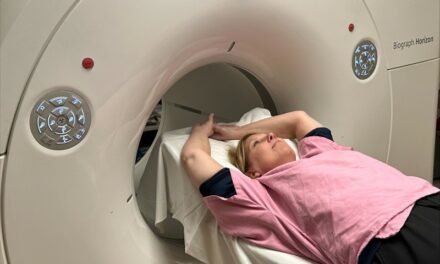
Total body PET/CT scans can successfully visualize systemic joint involvement in patients with autoimmune arthritis, according to new first-in-human research published in the October issue of The Journal of Nuclear Medicine. The total body PET/CT scans showed high agreement with standard joint-by-joint rheumatological evaluation and a moderate to strong correlation with rheumatological outcome measures.
Autoimmune inflammatory arthritides (AIA)—such as psoriatic arthritis and rheumatoid arthritis—are chronic, systemic conditions that cause joint inflammation, joint destruction and pain. According to the Centers for Disease Control and Prevention, approximately one in four adults, or 58 million Americans, have been diagnosed with arthritis by a doctor. By 2040, an estimated 78 million adults are projected to be diagnosed with arthritis.
“Currently, there are significant clinical challenges in managing AIA populations. For example, it is unclear which patients should receive which treatments, how exactly these treatments change the inflammatory status of different tissues or outcomes, and the impact the disease and treatments have on other organs of the body,” says Abhijit J. Chaudhari, PhD, professor of radiology at the University of California–Davis. “Systemic molecular imaging enabled by total body PET could provide currently unavailable, objective biomarkers that could help address these challenges.”
To assess the performance of molecular imaging to evaluate AIA, researchers used an ultra-low dose 18F-FDG total-body PET/CT acquisition protocol to scan 30 participants (24 with AIA and six with osteoarthritis). Participants also underwent a joint-by-joint rheumatological evaluation. In total, 1,997 joints were evaluated.
Total body PET/CT was successful in visualizing 18F-FDG uptake at joints throughout the entire body, including those of the hands and feet, without the need for custom positioning of the participants. In the AIA cohort, there was concordance between total body PET qualitative assessments and joint-by-joint rheumatological evaluation for 69.9% of the participants. About 20% of the AIA group had joints that were deemed negative on rheumatological evaluation but were positive on PET/CT, and 10% were negative on PET/CT but positive on the rheumatological evaluation.
For the osteoarthritis cohort, there was agreement in joint assessment among 91.1% of participants. In this group 8.8% of joints were deemed negative on rheumatological evaluation but were positive on PET/CT, and no joints were negative on PET/CT but positive on rheumatological exam.
“Systemic evaluation of arthritic disease activity across all musculoskeletal tissues of the body may provide unique insights for assessing disease burdenrisk stratification, treatment selection, and monitoring on treatment response. The biomarkers imaged may also have clear potential to accelerate arthritic drug discovery and development,” notes Chaudhari. “In addition, the inflammatory arthritides assessed in the paper are part of a broad category of autoimmune disorders. This work may contribute to improved understanding of total-body impact of autoimmunity.”


