Richard Semelka, MD Richard Semelka, MD |
Whole-body MRI is a fast, reliable, safe and accurate means of detecting disease throughout the entire body. This has been well shown in a recent article, 1 in which the authors demonstrated that a screening MR protocol showed various disease processes with accuracy almost equal to that of a variety of comparison “gold standard” diagnostic tests. That article emphasized one component of whole-body MRI, which is the use of gadolinium-enhanced 3D gradient echo imaging. There are, however, five major technical advances that have rendered whole-body screening with MRI a viable method, which include:
- remote movement of the imaging table from the imaging console
- multiple input channels that allow simultaneous use of multiple localized, specialized surface coils that generate high-resolution (high image quality) images of multiple regions of the body, without the delay of coil exchange and setup
- specialized surface coils that are designed for independent operation of the individual coil elements
- concurrent development of sequence (data acquisition) technology that operates in conjunction with the new specialized coils, termed parallel imaging
- development of high image quality 3D T1-weighted gradient echo (a type of MR data collection) with short echo time (TE) that facilitates acceptable imaging quality of various organ systems, most notably the lungs.
NEW CAPABILITIES
The combined effect of all these innovations has allowed, among other things: full body imaging to be performed more rapidly (because of remote table movement and new short data acquisitions), with maintenance of high image quality (simultaneous use of multiple specialized coils), and with the ability to image the lungs with adequate image quality (3D T1-weighted gradient echo imaging). The result is that the entire body can be imaged in a matter of 10 to 15 minutes, with high image quality maintained.
For an imaging study to be used to screen for disease, it must be able to detect disease accurately and avoid describing normal parts of the body as diseased. Since whole-body CT and MRI screening are relatively new procedures, limited data have been collected comparing the two methods. There is growing concern in the medical community that whole-body CT screening leads to a large number of questionable findings, requiring additional procedures, including surgery, creating added risks and costs for the individual. 2 Most of the information comparing the performance of CT and MRI has been gathered through diagnostic studies, that is, imaging studies ordered for the detection of suspected disease. Overall, MRI has been found to discover more lesions and correctly characterize disease, whether benign or malignant, than CT. In these capacities, MRI has been shown to be superior to CT for examining specific regions of the body, for example, head, abdomen, and pelvis. 3-8 In the past, the sole exception to this rule has been imaging of the lungs, which is performed better by CT than MRI, although new MRI techniques show promising results. 1,9
COST CONSIDERATIONS
 Figure 1. Liver. A: Coronal T2-weighted single shot echo-train spin-echo image. There is a high signal intensity lesion on T2-weighted image consistent with biliary hamartoma (arrow). B: Transverse T1-weighted fat suppressed 3D gradient echo image. Biliary hamartoma shows low signal intensity on post gadolinium T1-weighted image (arrows). Figure 1. Liver. A: Coronal T2-weighted single shot echo-train spin-echo image. There is a high signal intensity lesion on T2-weighted image consistent with biliary hamartoma (arrow). B: Transverse T1-weighted fat suppressed 3D gradient echo image. Biliary hamartoma shows low signal intensity on post gadolinium T1-weighted image (arrows). |
Cost of the imaging study is also an important consideration. Because of the nature of the complexity of the imaging system and intrinsic maintenance costs, MRI is unavoidably a more expensive test than CT. The machinery has many more components, and the requirement for liquid helium and liquid nitrogen to be continuously replenished renders it intrinsically more expensive. The real cost of MRI is greater than that of CT; an approximate estimate is that studies are about 20% more expensive.
MRI also is a safer modality than CT, due to both the imaging system itself and the intravenous contrast agent employed. 10-14 The powerful magnetic field and radiofrequency energy of MRI have not been shown to cause cancer or fetal abnormalities, unlike the ionizing radiation used in CT. It is important to note that although x-rays are known to cause cancer, the exact risk of cancer from receiving CT scans, and even repeat CT examinations is unknown. A more in-depth discussion on cancer risks from radiation exposure in CT and other x-ray procedures has been recently published. 15 The intravenous gadolinium-containing contrast agents used routinely in MRI are also considerably safer than the analogous intravenous agents employed in contrast-enhanced CT, which are iodine-based agents. There is a reduced association with kidney injury with the MRI contrast agents, and a reduced association with allergic reactions, including severe allergic reactions that can lead to death. 16-18 That is why, in general, individuals who are undergoing diagnostic imaging studies will have an MRI study rather than CT if they have poor kidney function or a history of allergies. Another very compelling aspect of the use of intravenous contrast agents with MRI is that the diameter of the needle inserted is much smaller with MRI, the volume of contrast material that is injected intravenously is 10 times less than with CT (the rapid injection of the large volume of contrast with CT can cause nausea), and the injection rate is slower with MRI than with CT. Additionally, the chance of the injected contrast agent not going into the vein but into the surrounding tissues is also greater with CT, which is a problem compounded by the large volume of the fluid injected.
LIMITING FACTORS
 Figure 2. Chest. A: Coronal T1-weighted fat suppressed 3D gradient echo post gadolinium image. B: Transverse T1-weighted fat suppressed 3D gradient echo post gadolinium image. Figure 2. Chest. A: Coronal T1-weighted fat suppressed 3D gradient echo post gadolinium image. B: Transverse T1-weighted fat suppressed 3D gradient echo post gadolinium image. |
Therefore, if MRI is superior to CT for finding small volume disease, correctly classifying disease, demonstrating that normal structures are intact and not diseased, safer, and less painful (smaller needlestick with MRI), then why is MRI still not considered the method of choice for whole-body screening? Historically, the primary reason for this is that MRI is much slower to perform than CT, and imaging of the lungs was suboptimal. Over the recent few months, the MR systems that are manufactured have imaging capability that allows them to image the entire body much faster while maintaining high image quality that even machines made 1 year ago did not possess. Basically, new designs of transmit-receiver coils, easier movement of the imaging table, and new data acquisition techniques have allowed rapid imaging of the entire body, and also acceptable image quality of the lungs. High-quality, whole-body MR imaging that would take 2 hours even 1 year ago now can be done in 10 to 15 minutes, making the technique very suitable for rapid, highly accurate whole-body imaging in an easily tolerable time frame. Other reasons for employing CT include: cost, the number of available systems (greater with CT), MRI studies are more difficult to perform (because there are more different types of data acquired), the interpretation of MR images is more complex, and fewer radiologists are trained to interpret MRI than CT.
 Figure 3. Pelvis. A: Transverse T2-weighted single shot echo-train spin-echo image. B: Transverse T1-weighted fat suppressed 3D gradient echo post gadolinium image. Figure 3. Pelvis. A: Transverse T2-weighted single shot echo-train spin-echo image. B: Transverse T1-weighted fat suppressed 3D gradient echo post gadolinium image. |
An important consideration when performing a screening study is what to look for. Unfortunately, even with whole-body imaging, every single disease process cannot be looked for in great detail. This is due to the fact that very detailed imaging of every part of the body would take an extremely long time even on the newest MRI machines. Therefore, studies should be tailored to look for the most common lethal diseases that afflict the general population in the part of the world the study is performed and also tailored to the types of disease that the person in particular is at risk of acquiring. The most prevalent diseases in North America include stroke, heart attack, and the common deadly cancers: lung, breast (in women), colon, prostate (in men), pancreas, lymphoma, and liver. Screening for common diseases in the general public should be supplemented with screening for disease that the particular person undergoing the screening study is at risk for (for example, kidney cancer in patients with kidney failure). I foresee the day, in the not too distant future, when, based on a computer-generated reading of genetic material obtained from a blood sample, doctors will know what diseases we have a propensity to acquire, and then the MRI scan will be performed specifically looking for those diseases.
 Figure 4. Brain. A: Transverse T2-weighted fat suppressed single shot echo-train spin-echo image. B: Transverse T1-weighted fat suppressed 3D gradient echo post gadolinium image. Figure 4. Brain. A: Transverse T2-weighted fat suppressed single shot echo-train spin-echo image. B: Transverse T1-weighted fat suppressed 3D gradient echo post gadolinium image. |
Although at present, reliability and accuracy of MR imaging at detecting diseases of certain organs including the liver, brain, spine, pancreas, and kidneys are extremely high and imaging of the lungs is reasonable, optimal imaging of the heart, breast, and colon still requires further development to achieve consistent image quality and consistent accurate display of small volume disease. Nonetheless, imaging of all the latter mentioned organs is currently at a diagnostically acceptable level.
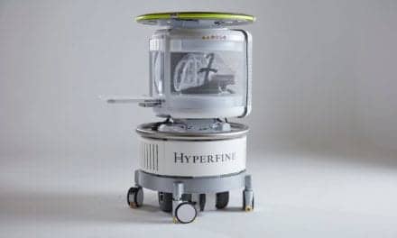
Currently manufactured MRI systems have evolved to the point that the equipment can image through the entire body with good image quality and sensitivity to detect disease within a 15-minute period. Whole-body, high-quality MRI screening is now feasible.
Richard Semelka, MD, is professor of radiology, director of MR services, and vice chair of clinical research, Department of Radiology, University of North Carolina, Chapel Hill.
References:
- Lauenstein TC, Goehde SC, Herborn CU, et al. Whole-body MR imaging: evaluation of patients for metastases. Radiology. 2004;233:139-148.
- Ko J, Casola G. Whole body MR screening found feasible. Whole body CT screening. Available at: www.rsna.org/publications/rsnanews/febo4/mri-1.html.
- Gillman S. Imaging the brain: second of two parts. N Engl J Med. 1998;338:889-896.
- Semelka R, Martin D, Balci C, Lance T. Focal liver lesions: comparison of dual-phase CT and multisequence multiplanar MR imaging including dynamic gadolinium enhancement. J Magn Reson Imaging. 2001;13:397-401.
- Low R, Semelka R, Woranwattanakul S, Alzate G, Sigeti J. Extrahepatic abdominal imaging in patients with malignancy: comparison of MR imaging and helical CT, with subsequent surgical correlation. Radiology. 1999;210:625-632.
- Low R, Semelka R, Woranwattanakul S, Alzate G. Extrahepatic abdominal imaging in patients with malignancy: comparison of MR imaging and helical CT in 164 patients. J Magn Reson Imaging. 2000;12:269-277.
- Semelka RC, Kelekis NL, Molina PL, Sharp TJ, Calvo B. Pancreatic masses with inconclusive findings on spiral CT: is there a role for MRI? J Magn Reson Imaging. 1996;6:585-588.
- Sheridan MB, Ward J, Guthrie JA, et al. Dynamic contrast-enhanced MR imaging and dual-phase helical CT in the preoperative assessment of suspected pancreatic cancer: a comparative study with receiver operating characteristic analysis. AJR Am J Roentgenol. 1999;173:583-90.
- Bader T, Semelka R, Pedro M, Armao D, Brown M, Molina P. Magnetic resonance imaging of pulmonary parenchymal disease using a modified breath-hold 3D gradient-echo technique: initial observations. J Magn Reson Imaging. 2002;15:31-38.
- Budinger TF. Nuclear magnetic resonance (NMR) in vivo studies: known thresholds for health effects. J Comp Assist Tomogr. 1981;5:800-811.
- Reid A, Smith FW, Hutchison JM. Nuclear magnetic resonance imaging and its safety implications: follow-up of 181 patients. Br J Radiol. 1982;55:784-786.
- McRobbie D, Foster MA. Pulsed magnetic field exposure during pregnancy and implications for NMR fetal imaging: a study with mice. Magn Reson Imaging. 1985;3:231-234.
- Heinrichs WL, Fong P, Flannery M, et al. Midgestational exposure of pregnant BALB/c mice to magnetic resonance imaging conditions. Magn Reson Imaging. 1988;6:305-313.
- Terens WL, Gluck R, Golimbu M, Rofsky NM. Use of gadolinium-DTPA-enhancement MRI to characterize renal masses in patient with renal insufficiency. Urology. 1992;40:152-154.
- Berrington de González A, Darby S. Risk of cancer from diagnostic X-rays: estimates for the UK and 14 other countries. Lancet. 2004;363:345-51.
- Brezis M, Epstein FH. A closer look at radiocontrast-induced nephropathy. N Engl J Med. 1989;320:179-181.
- Berns A. Nephrotoxicity of contrast media. Kidney Int. 1989;35:730-740.
- Parfrey PS, Griffiths SM, Barrett BJ, et al. Contrast material-induced renal failure in patients with diabetes mellitus, renal insufficiency, or both. A prospective controlled study. N Engl J Med. 1989;323:143-149.